Rajasthan Board RBSE Class 11 Biology Chapter 36 Cockroach
RBSE Class 11 Biology Chapter 36 Multiple Choice Objective Questions
Question 1.
Cockroach is
a. Herbivorous
b. Carnivorous
c. Omnivorous
d. Frugivorous
Question 2.
The head of cockroach is formed by the fusion of how many segments?
a. 5
b. 6
c. 7
d. 8
Question 3.
Number of segments in the embryonic abdomen
a. 10
b. 7
c. 11
d. 9
Question 4.
Exoskeleton of cockroach is made up of
a. Chitin
b. Cartilages
c. Bones
d. Keratin
Question 5.
The Johnston organ found in the antennae are
a. Gustoreceptors
b. Olfactoreceptors
c. Phonoreceptors
d. Motion Receptor
Question 6.
The mouth part is cockroach that functions like tongue
a. Labium
b. Hypopharymx
c. Mandible
d. Maxilla
Question 7.
Anal styles are found
a. Only in male cockroach
b. Only in female cockroach
c. Both
d. None
Question 8.
The body cavity of cockroach is
a. Coelom
b. Blastocoel
c. Haemocoel
d. Lvmphocoel
Question 9.
Number of cuticular teeth in the gizzard of cockroach
a. 4
b. 6
c. 8
d. 10
Question 10.
Hind gut in cockroach originates from
a. Ectoderm
b. Endoderm
c. Mesoderm
d. All the above
Question 11.
Number of spiracles in cockroach
a. 12 pairs
b. 14 pairs
c. 10 pairs
d. 20 pairs
Question 12.
The blood in cockroach is called is
a. Lymph
b. Haemolymph
c. Haemoglobin
d. None
Question 13.
Number of nerves in the peripheral nervous system of cockroach
a. 10 pairs
b. 20 pairs
c. 30 pairs
d. 40 pairs
Question 14.
The main excretory substance in cockroach is
a.Urea
b. Ammonia
c. Amino acid
d. Uric acid
Question 15.
The excretory organs in cockroach
a. Malpighii tubules
b. Fat body cells
c. Uricose glands
d. All the above
Question 16.
The hormone ecdysone which controls ecdysis is secreted by
a. Corphora allata
b. Neurosecretory cells
c. Prothoracic glands
d. Brain
Question 17.
The structural & functional unit of compound eye in cockroach
a. Ommatidium
b. Cornea
c. Retina
d. Rhabdone
Question 18.
Which of the following is a gland of female reproductive system
a. Phallic glad
b. Mushroom gland
c. Colleterial gland
d. Uricose gland
Question 19.
Number of eggs in the ootheca of cockroach
a. 14
b. 16
c. 18
d. 20
Question 20.
The young cockroach is called as
a. Nymph
b. Maggot
c. Periblastula
d. Imago
Answers :
1. c
2. b
3. c
4. a
5. d
6. b
7. a
8. c
9. b
10. a
11. c
12. b
13. c
14. d
15. d
16. c
17. a
18. c
19. b
20. a
RBSE Class 11 Biology Chapter 36 Very Short Answer Questions
Question 1.
Write the names of species of cockroach found in India.
Answer :
Periplanata americana, Blata orientalis, Blatella germinica
Question 2.
Give the number of segments in cockroach during embryonic & adult stages.
Answer :
20 & 19
Question 3.
Where the Johanston organs are found? Give their function.
Answer :
Antennae, Motion receptor
Question 4.
Which type of mouth parts are found in Cockroach?
Answer :
Biting & Chewing type
Question 5.
Give the role of anal cerci found in the cockroach.
Answer :
Phonoreceptors
Question 6.
What is the number of legs in cockroach and how many segments are found in each leg.
Answer :
3 pairs; total 9 segments of 5 types.
Question 7.
Why the body cavity of cockroach is not called as true coelom?
Answer :
It is not lined by the mesodenn.
Question 8.
Give the food of cockroach?
Answer :
All types of food (omnivorous)
Question 9.
Name the elastic plate covering the spiracle in cockraoch.
Answer :
Peretreme
Question 10.
What is the number of spiracles in cockroach.
Answer :
3
Question 11.
What is the number of ganglia in the ventral nerve cord?
Answer :
9 ganglia (3 + 6)
Question 12.
Write the main excretory organ in cockroach.
Answer :
Malpighian tubules
Question 13.
How many ommatidia are found in each compound eye of cockroach?
Answer :
2000
Question 14.
What is the role of phallic gland in the cockroach?
Answer :
It secretes outermost covering of the spermatophore.
Question 15.
How many times cockroach moults during metamorphosis?
Answer :
7 to 10 times.
RBSE Class 11 Biology Chapter 36 Short Answer Questions
Question 1.
Describe the structure of head in cockroach.
Answer :
The head is attached to the thorax at 90° with the help of a thin & elastic neck. It is triangular & pear shaped. The head in cockroach is hypognathus. It is formed by the fusion of 6 segments. One pair of compound eyes are situated on the head which are black & kidney-shaped.
Question 2.
Describe in brief the mouth parts in cockroach.
Answer :
The cockroach has biting & chewing mouth parts which are also called as mandibulated mouth parts. The mouth parts are attached around the mouth ventral to the head. The mouth parts enclose a cavity in front of the mouth which is called as pre-oral cavity. It is divisible into two parts viz.—
- Salivarium
- Cibarium
Question 3.
Differentiate male & female cockroach.
Answer :
|
Male Cockroach |
Female Cockroach |
| (a) The body is comparatively short. | (a) The body is comparatively long. |
| (b) The abdomen is narrow and 9 segments are visible in it. | (b) The abdomen is comparative broad and 7 segments are visible in it. |
| (c) The 7th sternite is not boat shaped | (c) The 7th sternite is large and boat shaped. |
| (d) The 9th sternite is attached with a pair of anal styles. | (d) The anal styles are absent. |
| (e) The wings are large and extend beyond the abdomen. | (e) The wings are comparatively short and remain up to the abdomen. |
Question 4.
Write the salient features of wings in cockroach.
Answer :
The cockroach has two pairs of wings which are attached dorso-laterafly to die mesothorax & metathorax. First pair of wings are thick, hard & leathery. They are called as tegmina or elytra or coverites. The tegmina covers the 2nd pair of wings during rest. The second pair of wings are thin, transparent & membranous. During rest, they remain folded below the first pairs of wings. The wings have a network of veins containing haemolymph at the time of formation. Later on. the blood dries up & tire network of veins act as an endoskeleton. Now, they are called as nervures. Hie wings are formed as evagination of the body wall between the tergum & pleuron. Well developed wings have only two layers of the cuticle. The basal part of the each wing is attached to the sternum with the help of basilar muscle. There is a pair of tergostemal muscles in each thoracic segment which are attached between the tergum & the sternum and are also called as dorso-ventral muscles. The contraction & the relaxation of the basilar muscles and tergostemal muscles are responsible for the movement of the wings.
Question 5.
Draw a labelled diagram of alimentary canal.
Answer :
Question 6.
Explain in brief the process of respiration in cockroach.
Answer :
Respiration in cockroach is performed by the contraction & the relaxation of the tergosternal muscles. As a result of the contraction of the tergosternal muscles, the air squeezed out which is called as expiration. The relaxation of the tergosternal muscles reduces the pressure on the trachea, as air result the air rushes into the body through the spiracles. It is called as inspiration. During rest the process of breathing in involuntary & during active phase, it a voluntary process. The exchange of gases takes place by diffusion between the trachiole fluid & tissue fluid.
Question 7.
Draw a labelled diagram of blood sinuses in cockroach.
Answer :
Question 8.
Explain in brief die central nervous system in cockroach.
Answer :
It includes nerve ring and the nerve cord.
Nerve ring :
It is situated in the head around the oesophagus and consists of a pair of supra-oesophageal ganglia, a pair of circum-oesophageal connectives & a pair of sub- oesophageal ganglia. The supra-oesophageal ganglia are also called as brain and is formed by the fusion of three pairs of ganglia. The brain is a bilobed structure which has three parts-
- Protocerebrum
- Deuterocerebrum
- Tritocerebrum
The sub-oesophageal ganglia are also formed by the fusion of three pairs of ganglia. They remain situated below the oesophagus.
Question 9.
Where the Malpighian tubule are situated in cockroach? How they function?
Answer :
Malpighian tubules :
There are 72 to 90 malpighian tubules which are attached in 6 groups of 12 to 15 at the junction between the midgut & hind gut. The malpighian tubules are long & narrow tubules which are yellow and unbranched. They rennan freely suspended in the heaemolymph. The length of the tubules is about 26 mm & the the diameter is 0.5 mm. The wall of the malpighian tubules is thick & is made up to 6 large cuboidal cells. The lumen is narrow & has ciliated or brush border lining. It is surround by a peritoneum.
Physiology :
Each malpighian tubule is divisible into 2 parts-
- Proximal half absorptive part.
- Distal half secretory part
The distal half part of the malpighian tubule take CO2. H2O, sodium urates & potassium urates from the haemolymph and secrete them into its lumen. In the lumen CO2, H2O. react to form H2CO3 which dissociate to form H+ & HCO3– Similarly, the sodium & potassium utrates ionized to form Na+, K+ & urate ions. These ions react together & finally form uric acid, sodium bicarbonate & potassium bicarbonate.
Question 10.
Write short notes on
- Apposition image
- Superposition image
Answer :
- Apposition image :
It is formed during moderate to bright light. It is also called as mosiac image.
The pigmented sheaths expand to make die ommatidium optically isolated in the bright light. The image of one part of the object is formed in one ommatidium. Hence, it is a distinct image. In cockroach, only mosiac vision is formed. - Superposition image :
It is formed in dim-light & in the night. There is contraction of the pigmented sheat. Hence the optical isolation is lost. It is an indistinct image. The image of one part of the object is formed in many adjacent ommatidia
Question 11.
Draw labelled diagram of male reproductive organs of cockroach.
Answer :
Question 12.
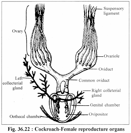
Draw labelled diagram of female reproductive organs in cockroach.
Answer :
Question 13.
Explain formation of ootheca in cockroach.
Answer :
Formation of Ootheca :
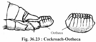
The ovipositors push the fertilized eggs into the oothecal chamber in 2 rows of 8 in each row. In the oothecal chamber. the secretions of the collateral glands form a dark brown colored covering around the fertilized eggs. The resulting structure is called as ootheca. The formation of ootheca takes about 20 hours.
The length of the ootheca is 8-10 mm. It’s one end is serrated. A female cockroach forms about 20-40 oothecae in it’s life span. The female carries the ootheca for few days & finally it deposit’s the ootheca in some dark, dry & warm place. The eggs of the cockroach are centrolecithal & are covered by chitinous shell. Every egg has a minute micropyle.
Question 14.
Explain structure of function of mushroom gland in cockroach.
Answer :
Mushroom or Utricular gland :
It is found in 7th abdominal segment & opens into an ejaculatory duct. It consists of three types of structures-
- Seminal Vesicles : They are small & round structures which store the sperms.
- Short central tubules or Utricular minores : They are small tubules which secrete nutrients for the sperms.
- Long peripheral tubules of Utriculi majors : They are long & thin tubules. Which secrete inner layer of the spermatophore.
Question 15.
Explain in brief the copulation & fertilization in cockroach.
Answer :
Copulation :
The copulation in cockroach takes place in the night in tail to tail position. It’s duration is of one hour. The copulation takes place during breeding season & the breeding season extends from March to September. During copulation, the male opens the female gonopore with the help of phallomeres and insert one spermatophore into the genital chamber. The spermatophore gets attached to the spermathecal papilla and the sperms get stored into the spermathecae. It takes about 20 hours. The empty spermatophore is forced out which is consumed as food by any cockroach. During breeding season, the male is attracted by the pheromone secreted by female cockroach.
Fertilization :
As a result of copulation. 16 ova are released from both the ovaries in the oothecal chamber. The ovipositors arrange the ova into two rows of 8-8 ova. Now the sperms are released on the ova from the spermathecae. Hence, the fertilization takes place in the genital chamber.
RBSE Class 11 Biology Chapter 36 Essay Type Questions
Question 1.
Describe in detail the morphology of cockroach.
Answer :
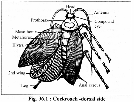
Shape, Size and Colour :
It’s body is dorso-ventrally flat, bilaterally symmetrical & dark brown in colour. Its length is 2 to 4 cm & width is 1 to 1.5 cm. There are two black spots on the dorsal side of the pronotum of the thorax.
Sexual dimorphism :
It exhibits distinct sexual dimorphism i.e. male and female can be distinguish on the basis of external features. These differences are as follows-
- Male cockroach
- Female cockroach
Segmentation :
The cockroach exhibits metameric segmentation. There are total 20 segments during embryonic stage but 19 segments during adult stage. The body is divisible into
head, thorax & abdomen.
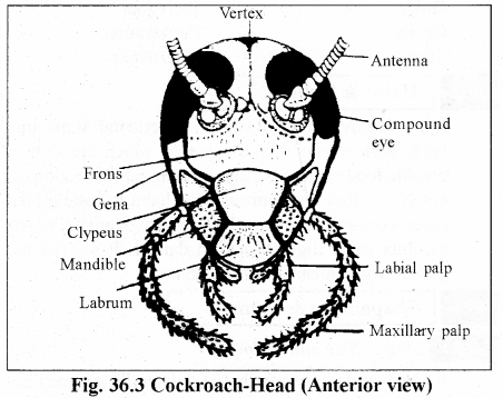
(A) Head –
The head is attached to the thorax at 90° with the help of a thin & elastic neck. It is triangular & pear shaped. The head in cockroach is hypognathus. It is formed by the fusion of 6 segments. One pair of compound eyes are situated on the head which are black & kidney-shaped.
There is a pair of simple eyes or ocellar spots or fenestra or ocelli which are situated near the compound eyes. They are non-functional & less developed. There is no image formation in the ocelli but they’ are sensitive to light. (Photosensitive)
The cockroach has a pair of antennae which are movable situated on the head in the antennary sockets near the compound eyes. Each antenna has three parts-
- Scape—Basal part which articulates to the head.
- Pedicel—Middle part which is provided with Jonston organ. The Johston organs are sensory to antennary movements.
- Flagullum—It is a thread-like & multi-segmented structure which is provided withe thigmoreceptors & olfactoreceptors.
The antennae of the cockroach are thin, long, filliform & monolifonn.
(B) Thorax –
It forms 2/5 part of the body and It has three segments viz.—prothorax, mesothorax & metathorax. The thorax has 3 pairs of legs and 2 pairs of wings. It bears 2 pairs of spiracles or stigmata which are situated ventrolaterally on pro-mesothorax & meso- metathorax.
(C) Abdomen –
It bears 11 segments during embryonic stage but 10 segments in adults. The abdomen is without locomotory organs. First eight abdominal segments bears 8 pairs of spiracles. The first abdominal segment is smallest.
There is a pair of scent glands between the 5th & 6th abdominal segments. Their secretions keep the enemies away. According to some scientists these secretions act as sex attractant.
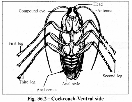
The abdomen bears a pair of anal cerci which are attached to the 10 th tergite in both male & female cockroaches. The anal cerci are long. thin. 15-segmented and provided withe phonoreceptors.
Male cockroach has a pair of small & pointed anal styles which are attached to the 9th sternite. They are unsegmented and help in copulation.
The anus is found at the end of the 10th segment in both male & female which is bounded by four chitinous plates viz.—epiproct-1, hypoproct-1 & paraproct-2. These podial plates are considered as the remnants of the 11th abdominal segment.
Female gonopore is found on the ventral side of the 8th abdominal segment and the male gonopore is found ventrally in the 9 the abdominal segment.
Both the male & female gonopores are provided with chitinous plates which are called as gonopophysis. The gonopophvsis are called as external genetelia or phallomeres. The female gonopophysis are called as ovipositors. The female gonopore remains covered by two gynovalvular plates which are modified 7th sternite.
Skeleton :
The cockroach bears both exoskeleton & endoskeleton.
(A) Exoskeleton
The body of cockroach is covered by chitinous exoskeleton which is dark brown in colour. The chitin is an acetate of glycosamine. The exoskeleton of each segment is called as sclerite which consists of four plates viz.—
- Tergum—one. dorsal & large
- Sternum—one. ventral & small
- Pleura—two, lateral & smallest
The sclerites remain connected by a thin & elastic membrane which is called as articular or arthrodial membrane. The chitin is soluble in water but it imparts hard nature to the exoskeleton
(a) Exoskeleton of head :
Exoskeleton of the head is called as head capsule which is formed by the fusion of six sclerites. The head capsule has 6 chitinous plates. The top of the head capsule is called as vertex which is divisible into two epicranial plates with the help of an epicranial suture. There is a frons plate just below the vertex & in front of the head capsule. The frons is followed by an oval plate, the clypeus. Both the lateral sides of the head capsule are made up of one-one gena plates which are also called as cheek plates. The head capsule bears an occipital foraman.
(b) Exoskeleton of neck :
The neck is covered withe 4 chitinous cervical plates. Out of them two arc dorso-lateral plates & two are ventro-lateral plates.
(c) Exoskeleton of thorax :
The sclerites of prothorax, mesothorax and metathorax are respectively called as pronotum, mesonotun & metanotum. The pronotum is the largest sclerite.
(d) Exoskeleton of abdomen :
The abdomen has 10 sclerites which are comparatively thin. In male the 7th tergite covers the 8th tergite. Similarly, the 7th stemite covers the 8the stemite. In female, 7th tergite covers 8th & 9th tergites and 7th stemite covers 8th & 9th stemites. In both male & female cockroaches 10th tergite is large & bilobed posteriorly.
(B) Endo skeleton
At many places, the exoskeleton in pushes to form endo skeleton to provides site for the attachment of muscles. The head bears a tent-like endo skeleton which is called as tentorium. The tentorium has a forman which allows nerves to pass through it.
The dorsal in pushing of the exoskeleton in the thorax are called apodedmes or phragma. There are three phragma viz.—
- First phragma— Betweem pronotum & mesonotum.
- Second phragma— Between mesonotum & metanotum.
- Third phragma— Between metanotum & first abdominal segment.
The ventral apodemes in the thorax are called as furca which are Y shaped.
Mouth Parts :
The cockroach has biting & chewing mouth parts which are also called as mandibulated mouth parts. The mouth parts are attached around the mouth ventral to the head. The mouth parts enclose a cavity in front of the mouth which is called as pre-oral cavity. It is divisible into two parts viz.—
- Salivarium
- Cibarium
It has total 8 mouth-parts which are of 5 types viz.—
- Labrum or upper lip : It is a chitinous plate which is attached movable to the clypeus. It’s distal end is elevated and the inner surface is attached with gustatory hair.
- Mandibles : There is a pair of mandibles which arc attached laterally below the labrum. Each mandible is triangular and its base is broad. It is attached with the help of a abductor muscle & a abductor muscle. The inner end of the mandible is provided withe denticles which help in cutting & chewing of the food. The inner basal end of the mandible is a pad-like which is called as prostheca. The prostheca bears tangoreceptors.
- First maxillae : There is a pair of first maxillae which are attached laterally just below the mandibles. It’s basal part is called as cardo which is attached to the head capsule withe the help of muscles. The distal end of the cardo is attached to a stipes structure and the distal end of the stipes of attached with an inner lacinia & an outer galea. They are used to clean antennae & legs. The lacinia is also used to hold the food during feeding.
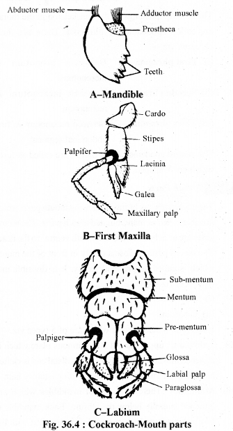
The outer surface of the stipes is attached with a maxillary palp which is situated on a palpifer plate. The maxillary palp is 5-jointed & it is provided with gustoreceptors.
It is formed by the fusion of one pair of second maxillae. Its proximal part is called as postmentum which is divisible into submentum & mentum. It’s distal part is called as prementum. A pair of palpiger plates are found on both the lateral sides of the prementum. There is a pair of labial palps which are 3-jointed & attached to the palpiger plates. The distal end of the prementum is attached with a pair of glossae (inner) and a pair of paraglossae (outer) The glossae & paraglossae help to keep the food particles in the pre-oral cavity. - Hypopharynx or Lingua :
It is a small, non-chitinous & cylendrical structure. It functions like a tongue and it is found attached to the common salivary duct in the preoral cavity.
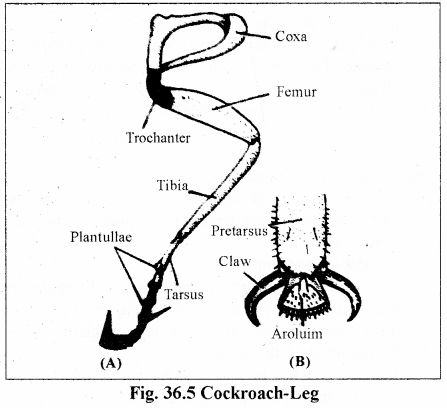
Legs :
The cockroach has 3 paris of jointed legs which are attached ventrolaterally to the thorax. Each leg consists of 9 segments which are of 5 types viz.—Coxa, Trochanter, Femur, Tibia & Tarsus.
The coxa is the proximal end which is attached movable to the thorax. The trochanter is the smallest part. The femur is the strongest part with is provided with bristles. The tibia is the longest part which is provided with tibial setae. The tibial setae are used to clean the body. The tarsus bears 5 segments which are called as tarsomeres or podomeres. Each tarsomere is attached with a pad like plantula. The last tarsomere is also called as pretarsus which is provided with 2 claws. The last plantula modifies to form an arolium or pulvilus which is attached between the claws. The arolium is a porous pad-like structure.
The claw’s help to locomote on rough surface and the arolium & plantullae help in locomotion on smooth surface.
Wings :
The cockroach has two pairs of wings which are attached dorso-laterafly to die mesothorax & metathorax. First pair of wings are thick, hard & leathery. They are called as tegmina or elytra or coverites. The tegmina covers the 2nd pair of wings during rest. The second pair of wings are thin, transparent & membranous. During rest, they remain folded below the first pairs of wings. The wings have a network of veins containing haemolymph at the time of formation. Later on. the blood dries up & tire network of veins act as an endoskeleton. Now, they are called as nervures. Hie wings are formed as evagination of the body wall between the tergum & pleuron. Well developed wings have only two layers of the cuticle. The basal part of the each wing is attached to the sternum with the help of basilar muscle. There is a pair of tergostemal muscles in each thoracic segment which are attached between the tergum & the sternum and are also called as dorso-ventral muscles. The contraction & the relaxation of the basilar muscles and tergostemal muscles are responsible for the movement of the wings.
Question 2.
Explain Digestive system in cockroach.
Answer :
Digestive system :
The digestive system includes alimentary canal & salivary glands.
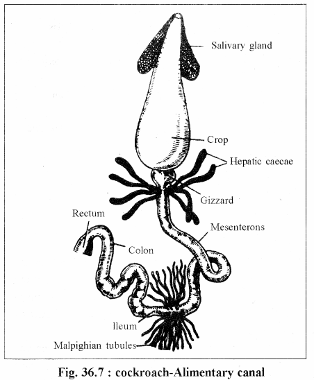
Alimentary canal :
It is a simple tubular structure which is divisible into three parts-
- Foregut or Stoinodaeum : It orgmates from the ectoderm & it is lined by the cuticle. It includes mouth, oesophagus, crop & gizzard.
- Mid gut or Mesenterone : It originates from the endoderm.
- Hind gut or Proctodaeum : It originates from the ectoderm & lined by the cuticle. It includes ileum, colon, rectum & anus.
The various parts of the alimentary canal are as follows –
- Mouth : It is situated at the base of the preoral cavity & it leads directly in to the oesophagus.
- Oesophagus : It is a narrow & simple tube which is found in the neck & the prothorx. It is folded internally.
- Crop : It is a bag-like structure which is thin-walled & elastic. It is found in the mcsothorax, metathorax & first 3 or 4 abdominal segments. It’s wall is renewable or breakable.
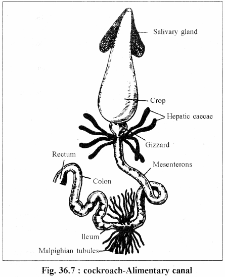
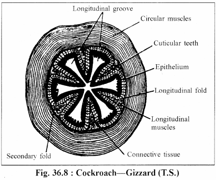
- Gizzard : It is also called as proventncuius. It is a small, thick walled, hard & conical structure which is found in the abdomen. It is surrounded by a thick layer of circular muscles.
It’s cavity is lines by the cuticle. Its anterior part is called as armarium which bears 6 chitinous & cuticular teeth. There are six longitudinal grooves alternate to the teeth which are provided with longitudinal muscles & bristles. These grooves function as strainer. The posterior part of the gizzard is funnel shaped. It has pads bearing bristles.
The posterior part extends into the mid-gut to form a stomodael valve. This valve prevents reverse entry of the food. - Mesenterone : It is the soft & delicate part of the alimentary canal which is 1/3 of the alimentary canal. Its anterior part is called as cardia which receives 6 to 8 hepatic caecae. The hepatic caecae are attached at the junction between the fore gut & mid gut.
The hepatic caecae are thin & blind tubes which are yellowish in color. The hepatic caecae secretes the digestive enzymes. The anterior half of the mid-gut is called as secretory part and the posterior half part as absorptive part. - Ileum : It is a small & narrow tube. It receives malpighian tubules which are attached at the joint between the mid gut & the hind gut. It is lined by the cuticle & internally it has many spines.
- Colon : It is a long & broad tube which is coiled. It is without spines.
- Rectum : It is the last part which opens outside through the anus It is small, broad & provided withe 6 longitudinal rectal papillae. The rectal papillae are padlike structures bounded by thin cuticle.
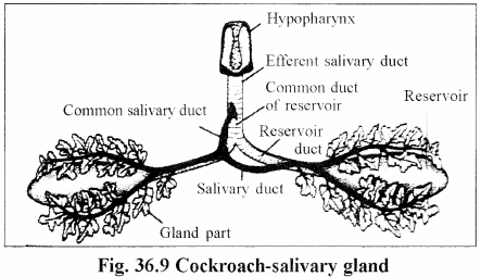
Salivary glands :
There is a pair of salivary glands which are situated in the thorax on both the sides of the crop. Each salivary gland is bipartite i.e. it’s anterior 1/3 part is bilobed. The salivary gland gives out a salivary duct. Both the salivary ducts joins to form a common salivary duct. The salivary glands secrete saliva.
There is pair of reservoirs which are called as receptacles. The reservoirs are the thin-walled & transparent bag-like structure which store the saliva. Each reservoir gives out a reservoir duct. Both the reservoir ducts join to form a common reservoir duct. The common salivary duct & the common reservoir duct join to from an efferent salivary duct which open into the pre-oral cavity.
Digestion :
It is an omnivorus animal which feeds with the help of biting & chewing mouth parts. In the pre-oral cavity, the food receives saliva with contains mucous and enzymes (amylase, chitinase & cellulase). The mucous makes the food slimy & soil.
The amylase, the chitinase & the cellulase perform part digestion of various carbohydrates. The partly digested food in the crop receives the hydrolases secreted by the hepatic caecae viz.—invertase. maltase lactase, trypsin, proteiase, lipase etc. They perform semi-digestion of the food. The semi-digested food enters into the gizzard where it is subjected to the grinding. The gizzard also filters the semi-digested fine food particles which enter into the mid-gut.
The stomodael valve secretes a peritrophic membrane around the semi-digested fine food particles. The peritrophic membrane is made up of protein & chitin. It is permeable to the digested food & the hydrolases. The peritrophic membrane protects the soft mid-gut from the hard food particles.
The secretory part of the mid-gut secretes the various hydrolases which complete the digestion of the food. Most of the digested food is absorbed by the secretory part of the mid gut. In the ileum, the spines tear the peritrophic membrane and absorbs the remaining digested food. The colon absorbs the water but most of the water is absorbed by the rectal papellae in the rectum. The fecal matter is digested in the form of small dry pellets.
Question 3.
Explain respiratory system in cockroach. Draw labelled diagram.
Answer :
Respiratory system :
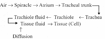
It exhibits aerial respiration which is with the help of tracheal system. The tracheal system carries the oxygen directly upto the tissues. It includes spiracles & trachea.
Spiracles :
It has 10 pairs of spiracles which are also called as stigmata. First two pairs of the spiracles are found vento-laterally in pro/mesothrax and meso/metathorax. The first pair of the abdominal spiracles are found dorsolaterally on the 1st abdomen segments. The seven pairs of the spiracles are found ventro-laterally in 2nd to 8th segment on the pleurites
Each spiracle is oval and it is surrounded by a chitinous ring called peritreme. The spiracles are provided with ciliary brislles which filter the inspired air. The thoracic spiracles are comparatively large. The first thorasic & the first abdominal spiracles remain permanently open. Rest spiracles open only at the time of inspiration and they are provided with valves. Each spiracle leads into a small chamber, tire tracheal chamber or atrium. Hence, there are 10 pairs of atria.
Trachea :
The trachea is also called as wind pipe & it originates from ectoderm. It has three pairs of longitudinal tracheal trunks
- Dorso-lateral tracheal trunk—1 pair
- Latral tracheal trunk—1 pair
- Ventro-latral tracheal trunk—1 pair
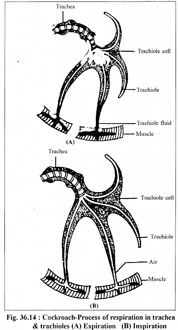
The 10 atria of one side open into the ventro-lateral tracheal tracheal trunks of it’s own side. All the six longitudinal tracheal trunks remain connected by transverse connecting trachea in each segment. The tracheal trunks divide to form trachea. The trachea are thin-walled, elastic & silvery’ white tubes. Their walls are supported by spiral rings made up of chitin. Each trachea ends into a trachiole cell. The trachiole cell gives out 3 to 5 intracellular tubules which are called as trachioles. The trachioles are the tubules of 1 micron diameter. They are silvery white & their walls are supported by spiral rings made up of trachin protein, the inner ends of the trachioles are closed & are full of trachiole fluid. This trachiole fluid helps in the exchange of the gases. The lining of the trachiole is called as intima.
Mechanism of Respiration :
Respiration in cockroach is performed by the contraction & the relaxation of the tergosternal muscles.
As a result of the contraction of the tergosternal muscles, the air squeezed out which is called as expiration. The relaxation of the tergosternal muscles reduces the pressure on the trachea, as air result the air rushes into the body through the spiracles. It is called as inspiration. During rest the process of breathing in involuntary & during active phase, it a voluntary process. The exchange of gases takes place by diffusion between the trachiole fluid & tissue fluid. The route of air during breathing as follows
Question 4.
Draw labelled diagram of heart in cockroach and explain its physiology.
Answer :
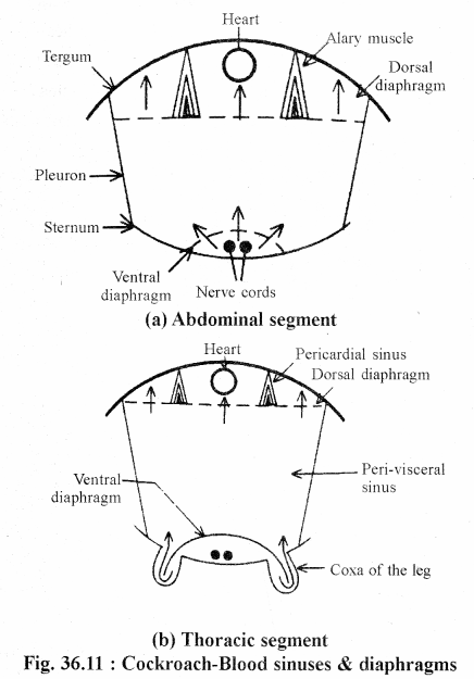
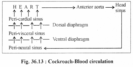
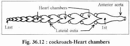
It has a tubular & pulsatile heart which is found mid- dorsally in the pericardial sinus. The heart has 13 chambers. Out of them 3 are found in the thorax & 10 are situated in the abdomen.The heart is closed posterior. Anterior, the heart opens into an anterior aorta which leads into the head sinus. Each heart chamber is averted funnel-shaped and it has two lateral
ostia. The blood form the pericardial sinus enters into the heart through these lateral ostia. In the heart the blood flows from behind to forwards. The first heart chamber is largest & the last heart chamber is smallest. The heart of the cockroach is neurogenic. The heart beats in cockroach are 49 per minute.
There are 13 pairs of alary muscles i.e. every segment of the thorax & abdomen has, a pair of alary muscles. The alary muscle is triangular. Its broad part is attached to the diaphragm & the pointed part to the tergum. They are also attached to the heart wall with the connective tissues. The alary muscles of the thorax are large. The alary muscles of the 10th abdominal segment.
In the cockroach, the blood circulates with the help of the alary muscles & the pulsatile heart. The contraction of the heart is called as systole & its relaxation as diastole. The duration between to heart beats is called as diastasis. As a result of the contraction alary-muscles, the blood of the perivisceral sinus enters into the pericardial sinus and finally into the heart through the lateral ostia.
Question 5.
Explain structure of compound eye in cockroach.
Answer :
Compound Eyes :
There is a pair of compound eyes which are situated on the head on near the sides of the vertex. The compound eyes are black, kidney-shaped and sessile. Each eye is covered by a transparent cornea which has 2000 hexagonal corneal facets.
Each corneal facet represents an ommatidium. Hence, there are 2000 ommatidia in each compound eye. The ommatidium is a structural & functional unit of the compount eye.
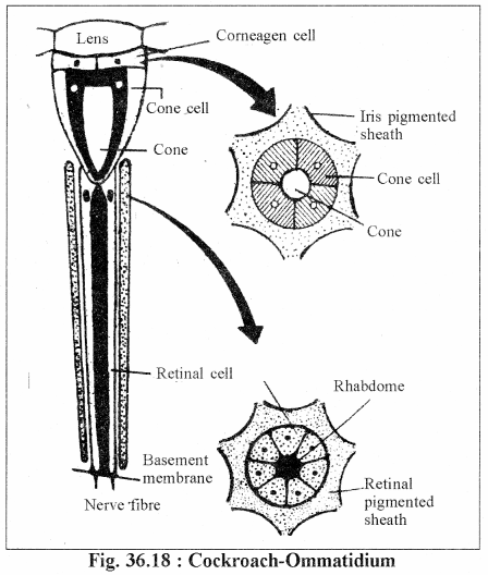
Structure of ommatidium :
The corneal facet is like a biconvex lens. This cornea! lens is secreted by two comeagen cells which are situated just below the facet. There are 4 cone cells below the comeagen cells which are also called as vitrelle. The cone cells secrete a crystalline cone which is found in the centre of the cone cells. There are seven retinal cells which ae situated around a rhabdome just below the cone cells. The rhabdome is secreted by the retinal cells. Each compound eye has 14,000 retinal cells which are situated on a basement membrane. Each retinal cell gives out a nerve fibre, hence, all the 14,000 nerve fibres unite to form an optic nerve.
The cornea, the comeagen cells & the cone cells together form dioptric part. Similarly, the rhabdome & the retinal cells together form receptor part.
The ommatidium remains optically isolated with the help of pigmented sheaths. The dioptric part is covered with retinal pigmented sheath. The pigmented sheaths of the cockroach are non-contractile. Hence, there is permanant optical isolation of the ommatidia. In other insects, the pigmented sheath are contractile & they contract in the dim-light. Hence, the optical isolation is lost during dim-light.
Working of the compound eye :
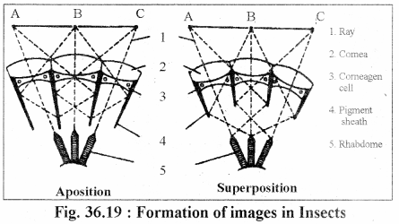
Normally, two types of images are formed in insects viz.
- Apposition image :
It is formed during moderate to bright light. It is also called as mosaic image.
The pigmented sheaths expand to make die ommatidium optically isolated in the bright light. The image of one part of the object is formed in one ommatidium. Hence, it is a distinct image. In cockroach, only mosaic vision is formed. - Superposition image
It is formed in dim-light & in the night. There is contraction of the pigmented sheet. Hence the optical isolation is lost. It is an indistinct image. The image of one part of the object is formed in many adjacent ommatidia
Question 6.
Explain male / female reproductive system in cockroach.
Answer :
Reproduction system :
The cockroach is a uni sexual animal & it exhibits sexual dimorphism.
Male reproductive organs :
The male cockroach has following reproductive organs-
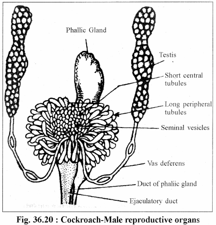
- Testes : There is a pair of testes in 4th. 5th & 6th abdominal segments. Each testis is tri-lobed. Each lobe has 20 tubular lobules.
- Vas deferens : Each testis leads into a vas deferens. Both the vas deferens open separately at the base of the mushroom gland.
- Mushroom or Utricular gland : It is found in 7th abdominal segment & opens into an ejaculatory duct. It consists of three types of structures—
(a) Seminal Vesicles : They are small & round structures which store the sperms.
(b) Short central tubules or Utricular minores: They are small tubules which secrete nutrients for the sperms.
(c) Long peripheral tubules of Utriculi majors : They are long & thin tubules. Which secrete inner layer of the spermatophore. - Ejaculatory duct : The male cockroach has a thin, glandular & contractile ejaculatory duct. It is attached to the ventral phallomere. It opens outside through the male gonopore. It secretes teh middle layer of the spermatophore.
- Phallic or Conglobate gland : It is a white & bag-like gland which is situated ventral to the ejaculatory duct from 6th to 9th abdominal segments. Its anterior end is broad & the posterior end is narrow tube-like. It opens outside through the male gonopore withe help of a duct. The duct is attached to the right

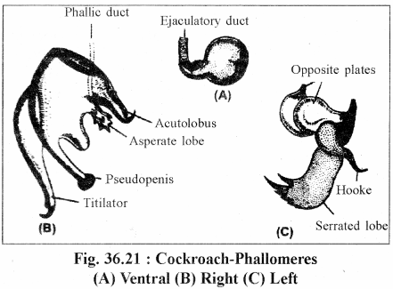
- Phallomeres of Gonapophyses :
The male gonopore is provided with three irregular chitinous plates which are called as phallomeres. They are as follows
(a) Left phallomere : It has 4 lobes viz.—asperate lobe, titilator. acutolobus & pseudopenis.
(b) Right phallomere : It includes a hook, a serrated lobe & two opposite lobes.
(c) Ventral phallomere : It is a small & flat plate. The ejaculatory duct is attached to it.
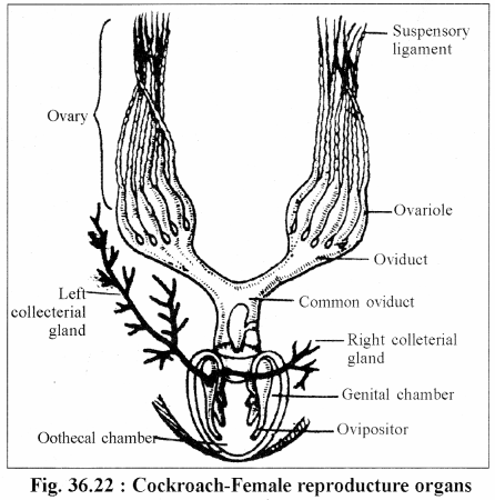
Female reproductive organs :
They female reproductive include –
- Ovaries :
There is a pair of ovaries which are found in 3rd to 6th abdominal segments. Each ovary has eight long, light yellow & tubular ovarioles.
Each ovariole has 5 parts –
(i) Suspensory ligament
(ii) Gennarium
(iii) Vitellarium
(iv) Ovarium or Egg chamber
(v) Pedicle
The suspensory ligaments of all the 8 ovarioles remain joined. - Oviduct & common oviduct :
All the eight ovarioles of one ovary open into an oviduct. Both the oviducts join to form a common oviduct or vagina which is found in 7th segment & opens into a genital chamber. - Gynatrium :
It is also called as brood pouch. It is formed by the modification of the 8th & 9th stemites. It is divisible into 2 parts—
(a) Genital chamber—Anterior & small
(b) Oothecal chamber—Posterior & large
The gynatrium opens outside through the female gonopore. - Spermathecae :
There is a pair of spermathecae which are unequal i.e. right spermatheca is comparatively small. They open through a common duct into the genital chamber. The opening is situated on a spermathecae papilla. They store sperms after the copulation. - Colleterial glands :
There is a pair of colleterial glands which are shiny, branched & uneuqal. The left gland is large & more branched.
Both the glands open through a common duct into the oothecal chamber. The right gland secretes dihydroxy phenol & the left gland secretes a protein. Both these substances react to form a scleroprotein which is dark brown in colour. - Female gonopophysis :
It has 6 chitinous plates attached to the roof of the oothecal chamber which are called as ovipositors. The female gonopore is covered by two gynovalvular plates which arc large & oval. They are formed by the modification of 7th sternum.
Reproduction physiology :
(i) Formation of spermatophore
It is formed in the male reproductive organs. It is a bag-like structure which is three-layered. It is full of sperms. The inner layer is secreted by the long peripheral tubules of the mushroom gland, the middle layer is secreted by the ejaculatory duct & the outer layer is secreted by the phallic gland
(ii) Copulation
The copulation in cockroach takes place in the night in tail to tail position. It’s duration is of one hour. The copulation takes place during breeding season & the breeding season extends from March to September. During copulation, the male opens the female gonopore with the help of phallomeres and insert one spermatophore into the genital chamber. The spermatophore gets attached to the spermathecal papilla and the sperms get stored into the spermathecae. It takes about 20 hours. The empty spermatophore is forced out which is consumed as food by any cockroach. During breeding season, the male is attracted by the pheromone secreted by female cockroach.
(iii) Fertilization
As a result of copulation. 16 ova are released from both the ovaries in the oothecal chamber. The ovipositors arrange the ova into two rows of 8-8 ova. Now the sperms are released on the ova from the spermathecae. Hence, the fertilization takes place in the genital chamber.
(iv) Formation of Ootheca
The ovipositors push the fertilized eggs into the oothecal chamber in 2 rows of 8 in each row. In the oothecal chamber. the secretions of the collecterial glands form a dark brown colored covering around the fertilized eggs. The resulting structure is called as ootheca. The formation of ootheca takes about 20 hours.
The length of the ootheca is 8-10 mm. It’s one end is serrated. A female cockroach forms about 20-40 oothecae in it’s life span. The female carries the ootheca for few days & finally it deposits the ootheca in some dark, dry & warm place. The eggs of the cockroach are centrolecithal & are covered by chitinous shell. Every egg has a minute micropyle.