Rajasthan Board RBSE Class 11 Biology Chapter 9 Structure and Functions of Cell Organelles
RBSE Class 11 Biology Chapter 9 Multiple Choice Objective Questions
Question 1.
The elaioplasts store –
(a) Starch
(b) Protein
(c) Glycogen
(d) Lipids
Question 2.
Suicidal bags of cell –
(a) Mictochondria
(b) Lysosome
(c) Dictyosome
(d) Plastid
Question 3.
The ribosomes found in mitochondria & chloroplasts-
(a) 70S
(b) 80S
(c) 50S
(d) 60S
Question 4.
The orgenelle which is not found in the plant cells-
(a) Plastid
(b) Microtubules
(c) Sphersomes
(d) Centrosome
Question 5.
The orgenelle which is not found in the animal cells-
(a) Plastids
(b) Microtubules
(c) Lysosomes
(d) Centrosome
Answers:
(1) d, (2) b, (3) c, (4) d, (5) a,
RBSE Class 11 Biology Chapter 9 Very Short Answer Questions
Question 1.
Altmann assign which name to mitochondria ?
Answer:
Bioplast.
Question 2.
The power house of the cell is?
Answer:
Mitochondria.
Question 3.
Write the names of leucoplasts ?
Answer:
Amyloplast, Elaiplast &Aleuroplast.
Question 4.
Name the components of Golgi body ?
Answer:
Lamillae, vacuoles & vesicles.
Question 5.
Name the structures of ER ?
Answer:
Cistemae, vesicles & tubules.
Question 6.
Name the two subunits of 70 S ribosome ?
Answer:
50 S & 30 S.
Question 7.
Who discovered centrosome ?
Answer:
Von Benden.
Question 8.
Which orgenelle performs intracellular digestion ?
Answer:
Lysosome.
RBSE Class 11 Biology Chapter 9 Short Answer Questions
Question 1.
Write the functions of mitochondria.
Answer:
Mitochondria are called as “Power house” of the cell. They peform redox reactions to release electrons, they pass the electrons through ETS to from ATP and they store ATP in the matrix.
Question 2.
Write the names of various types of plastids.
Answer:
The plastids may be pigmented or non-pigmented, on this basis, they are of three types (Schimper, 1983)
(i) Leucoplasts (ii) Chromoplasts & (iii) Chloroplasts
Question 3.
Give the names and one function of orgenelles bounded by single unit membrane.
Answer:
- ER – Skeletal support to the cell.
- Golgi body – It forms Lysosomes.
- Lysosome – It performs digestion.
- Spherosomes – It performs digestion.
Question 4.
What are microbodies ?
Answer:
They are minute bodies which are bounded by single unit membrane. They are formed by ER and are of three types-Spherosomes:
They are small spherical orgenelles found in the cytoplasm of plant cells. They are bounded by single membrane of lipoprotein. They are not found in animal cells. They are formed by ER. They synthesize and store lipids. Examples – Cells of cotyledons of mustard, groundnut, sesame etc. They are also called as oleosome. They were discovered by Perner (1953). Term sphaerosome was given by Dangeard (1919). Their diameter is about 0.5 to 2.5 micron.
Peroxisomes:
They were discovered by C. de Duve (1966-67). They are round & small orgenelles which are virtually found in all eukaryotes (All plant cells, liver & kidney cells of animals). They are abundance in photosynthetic cells of plants doing photo respiration. They are full of enzymes like oxidases, catalases which function like lysosome.
Functions :
- A major function of peroxisome is the breakdown of very long chain fatty acids.
- The process of photo respiration occurs jointly in peroxeosomes, chloroplasts & mitochondria.
- In animal cells, they perform fat metabolism.
- Their catalase enzyme degrades H2O2 (hydrogen peroxide) released in metabolic activities.
- They minimize the effects of toxins in the cytoplasm.
Glycoxysomes:
- They were discovered by Harry Beevers (1961). Normally, they are found in fat rich plant cells such as fat storage tissues of germinating seeds.
- Their enzymes initiate the breakdown of fatty acids to produce sugars (carbohydrate) by gluconeogenesis.
- In glycoxysomes, the fatty acids are oxidized to acetyl- COA by peroxisomal p-oxidation enzymes.
- They are absent in animal cells. They are minute orgenelles bounded by single unit membrane and perform glyoxylate cycle.
Question 5.
What is power house of cell? Write its origin and distribution.
Answer:
Mitochondria [Singular – Mitochondrion]
[Gr. Mitos = thread; Chondrion – granule]
They were first seen by Kolliker (1880) in the striated muscles of the insects. Fleming (1882) named them as “Fila”. Altmann (1890) called mitochondria as “Bioplast”. Term “mitochondria” was coined by C. Benda (1897-98). F. Meves (190) made the first recorded observation of mitochondria in plant cells (white water lilly).
Number:
They are found in all types of cells except the prokaryotic cells. They are round or rod-like or filamentous. The average length is 5 to 8 micron and the diameter is 0.5 to 1 micron. Their number varies from species to species and it is 50 to 5000. However, Microsterias green alga & sporozoite (a stage of Plasmoduim), its number is one. There are about 5 lakh mitochondria in Pelomyxa.
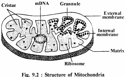
Structure:
They are bounded by two unit membranes viz. – Outer and inner membranes which are made up of proteins & phospholipids. The inner membrane forms numerous finger like processes into the cavity called Cristae. The cristae may be tubular (branched or unbranched) or vesicular. There is a perimitochondrial space between the outer and inner membranes which contains respiratory enzymes to perform oxidative phosphorylation. The central cavity of the mitochondria is full of matrix which consists of 70S ribosome, mDNA (mitochondrial DNA), all types of RNA, Redox enzymes and ATP.
The inner surface of inner membrane of cristae are attached with small granules which are called as elementary particles or respiratory granules or oxysomes. Every oxyosome consists of head, stalk and base. The diameter of head is about 86A°. These oxysomes remain equally spaced by 100A°. The oxysomes contain ATP synthetase enzyme which performs oxidative phosphorylation.
The mitochondrial DNA is circular and has more C-G combinations. The mitochondria shows auto reproduction ie. they divide to form new mitochondria. Therefore, they are also called as semiautonomous orgenelles. Chemically, they consists of proteins (65-70%), phospholipids (25%), RNA (0.5%); DNA & ATP etc.
Functions of Mitochondria:
- Mitochondria are called as “Power house” of the cell. They peform redox reactions to release electrons, they pass the electrons through ETS to from ATP and they store ATP in the matrix.
- They are the site of Krebs cycle, particularity the matrix.
- They are the site for oxidative phosphorylation, it is performed by oxyosomes.
- It is the main orgenelle that performs photorespiration.
Similarities between Plastids & Mitochondria:
- It is belived that both of them evolved from endosymbiotic bacteria.
- Both exhibit similar growth and origin.
- Both orgenelles contain DNA, RNA & 70S riboromes.
- Both are able to synthesize their own required point.
RBSE Class 11 Biology Chapter 9 Essay Type Questions
Question 1.
Given an account of history, structure and functions of mitochondria. Draw labelled diagram.
Answer:
Mitochondria [Singular – Mitochondrion]
[Gr. Mitos = thread; Chondrion – granule]
They were first seen by Kolliker (1880) in the striated muscles of the insects. Fleming (1882) named them as “Fila”. Altmann (1890) called mitochondria as “Bioplast”. Term “mitochondria” was coined by C. Benda (1897-98). F. Meves (190) made the first recorded observation of mitochondria in plant cells (white water lilly).
Number:
They are found in all types of cells except the prokaryotic cells. They are round or rod-like or filamentous. The average length is 5 to 8 micron and the diameter is 0.5 to 1 micron. Their number varies from species to species and it is 50 to 5000. However, Microsterias green alga & sporozoite (a stage of Plasmoduim), its number is one. There are about 5 lakh mitochondria in Pelomyxa.
Structure:
They are bounded by two unit membranes viz. – Outer and inner membranes which are made up of proteins & phospholipids. The inner membrane forms numerous finger like processes into the cavity called Cristae. The cristae may be tubular (branched or unbranched) or vesicular. There is a perimitochondrial space between the outer and inner membranes which contains respiratory enzymes to perform oxidative phosphorylation. The central cavity of the mitochondria is full of matrix which consists of 70S ribosome, mDNA (mitochondrial DNA), all types of RNA, Redox enzymes and ATP.
The inner surface of inner membrane of cristae are attached with small granules which are called as elementary particles or respiratory granules or oxysomes. Every oxyosome consists of head, stalk and base. The diameter of head is about 86A°. These oxysomes remain equally spaced by 100A°. The oxysomes contain ATP synthetase enzyme which performs oxidative phosphorylation.
The mitochondrial DNA is circular and has more C-G combinations. The mitochondria shows auto reproduction ie. they divide to form new mitochondria. Therefore, they are also called as semiautonomous orgenelles. Chemically, they consists of proteins (65-70%), phospholipids (25%), RNA (0.5%); DNA & ATP etc.

Functions of Mitochondria:
- Mitochondria are called as “Power house” of the cell. They peform redox reactions to release electrons, they pass the electrons through ETS to from ATP and they store ATP in the matrix.
- They are the site of Krebs cycle, particularity the matrix.
- They are the site for oxidative phosphorylation, it is performed by oxyosomes.
- It is the main orgenelle that performs photorespiration.
Similarities between Plastids & Mitochondria:
- It is belived that both of them evolved from endosymbiotic bacteria.
- Both exhibit similar growth and origin.
- Both orgenelles contain DNA, RNA & 70 S riboromes.
- Both are able to synthesize their own required point.
Question 2.
Describe the structure & function of chloroplasts found in higher plants with help of labelled diagram.
Answer:
Plastids were discovered and named by E.Haeckel (1865 – 1865). Schimper (1883-85) named the plastids as chioroplast which participate in photosynthesis. Plastids are bounded by two unit membranes and are found in the cytoplasm of eukaryotic plant cells. The chioroplasts are green due to chlorophyll and are slightly flat and round. They synthesize and store many bio-chemical substances. They are absent in animals, fungi & prokaryotic cells. The blue-green algae (cyanobacteria) have similar primitive structures called chromospheres.
Structure of Chloroplast:
They are bounded by two membranes made up of proteins and fats. Their thickness is 25 to 70A° and they have a broad inter-membranous space called periplastidial space. The dimensions of the chloroplasts is 5 to l0p x 2 to 4p.m. The inner jelly like fluid part of the chloroplast is called as stroma which consists of protein, starch, plastoglobuli, 70S ribosome, DNA and water etc.
The stroma consists of bundles of membranous lamellae called thylacoids. These thylakoids are arranged in stacks known as grana (Singular-granum). Every granum includes 2 to 100 thylacoids. The chlorophyll is found in the thylacoids and they are sites for the process of light reactions of photosynthesis. The thylacoids are interconnected by stroma lamellae. The inner surface of membranes of thylacoids are attached with minute granules called quantasome.
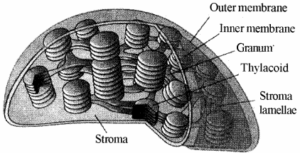
The name quantasome was given by Rodric B. Park and John Biggins (1994). Every quantasome consists of about 230 pigment molecules including photosynthetic pigments and redox carriers. The diameter of quantasome is 150 to 180A. The chemical composition of chloroplast is proteins (40 to 50%),’Lipids (23 to 25%), RNA (4%), DNA (0.03%), Chlorophyll (1 to 2%), carotinoids and trace elements like iron, zinc, manganese, magnesium, copper etc.
When the plants are kept in dark for longer period, the chloroplast form colourless etioplasts which contain lipids and volatile oils. These etioplasts reform chloroplasts when exposed to light.
Functions of Plastids:
- The chloroplasts synthesize carbohydrates food through photosynthesis. The light reaction takes place in grana and the dark reaction in the stroma.
- Leucoplasts help in storage of proteins, starch and oil. Such as amvloplasts, elaiplasts & aleuroplasts.
- Chloroplasts are also the site for photorespiration. The photorespiration needs chloroplasts, peroxysomes and mitochondria.
- They release oxygen by degrading water.
- They consume atmospheric COz and clean the
Question 3.
Give an account of microbodies found in cells.
Answer:
They are minute bodies which are bounded by single unit membrane. They are formed by ER and are of three types-
Spherosomes:
They are small spherical orgenelles found in the cytoplasm of plant cells. They are bounded by single membrane of lipoprotein. They are not found in animal cells. They are formed by ER. They synthesize and store lipids. Examples – Cells of cotyledons of mustard, groundnut, sesame etc. They are also called as oleosome. They were discovered by Perner (1953). Term sphaerosome was given by Dangeard (1919). Their diameter is about 0.5 to 2.5 micron.
Peroxisomes:
They were discovered by C. de Duve (1966-67). They are round & small orgenelles which are virtually found in all eukaryotes (All plant cells, liver & kidney cells of animals). They are abundance in photosynthetic cells of plants doing photo respiration. They are full of enzymes like oxidases, catalases which function like lysosome.
Functions :
(i) A major function of peroxisome is the breakdown of very long chain fatty acids.
(ii) The process of photo respiration occurs jointly in peroxeosomes, chloroplasts & mitochondria.
(iii) In animal cells, they perform fat metabolism.
(iv) Their catalase enzyme degrades H2O2(hydrogen peroxide) released in metabolic activities.
(v) They minimize the effects of toxins in the cytoplasm.
Glycoxysomes:
They were discovered by Harry Beevers (1961). Normally, they are found in fat rich plant cells such as fat storage tissues of germinating seeds.
Their enzymes initiate the breakdown of fatty acids to produce sugars (carbohydrate) by gluconeogenesis.
In glycoxysomes, the fatty acids are oxidized to acetyl- COA by peroxisomal p-oxidation enzymes.
They are absent in animal cells. They are minute orgenelles bounded by single unit membrane and perform glyoxylate cycle.
Question 4.
Give an account of structure & types of ribosomes. Explain union & dissociation of the sub units.
Answer:
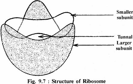
Robinson & Brown (1953) first discovered ribosomes in plant cell. In the animal cell, they were first observed by George Emil Palade (1955). The term ribosome was coined by R.B. Roberts (1958). They are oblate spheroidal in shape and they are not bounded by the membranes. Their dimensions are 150A° – 250A°. They are of two types viz. –
- Free ribosomes : They are found freely in groups in cytoplasm and inside the mitochondria & plastids.
- Bound ribosomes : They are attached the rough ER and outer nuclear membrane.
On the basis of sedimentation coefficient the ribosomes are of three types viz. –
- 70S ribosomes – It consists of two subunits of 50S & 30S. They are found in prokaryotes, mitochondria & plastids.
- 80S ribosomes – It consists of two subunits of 60S & 40S. They are found in eukaryotic cells.
- 55S ribosome – It consists of two subunits of 35S & 25 S. They are found in the mitochondria of mammalian cells.
The larger subunit is dome-shaped and the smaller unit is oval. The union and dissociation of these subunits depends upon mg++ concentration. Normally, these subunits are found separately. They unite to form ribosome only at the time of protein synthesis. During protein synthesis, the ribosomes unite to the mRNA to form a chain which is called as polyribosome or polyribosome or polysome. The ribosomes are mainly made up of RNA & protein and some Mg, Ca, Mn etc. The ribosomes participate in protein syntehsis.
Question 5.
Write short notes on-
(i) Centrosome
(ii) Vocuoles
(iii) Microtubules
Answer:
Vocuoles :
Dujardin (1941) discovered vacuoles in the plant cells. They may be one or more, small or large but always single membrane bounded round structures. The vacuoles that contains cell sap are called as sap vacuoles. They are bounded by a living membrane called tonoplast. In young cells, the vacuoles are small but their size increases with the increase in cell size. The cell sap indues sugar, amino acids, waste substances and mineral salts. They are called as Ergastic substances. The cell sap of fruits and flowers has dissolved anthocyanin pigments of blue, yellow, red etc. colours.
Types of Vacuoles:
On the basis of composition and functions, the vacuoles are of four types-
- Sap vacuoles – They store ergastic substances.
- Contractile vacuoles – They perform osmoregulation.
- Food vacuoles – Digestion of food takes place in these vacuoles.
- Air vacuoles – They are found in some prokaryotes. They store metabolic air & provide buyaney.
Functions of vacuoles:
The vacuoles perform following functions-
- They store and concentrate food materials and mineral ions.
- The help to maintain osmotic pressure and turgidity.
- They store waste products.
- Their anthocyanin provides attractive colours to the flowers & fruits that help in pollination of flowers & dispersal of fruits.
Centrosome:
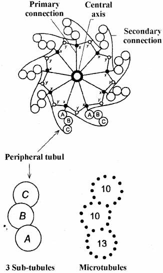
It was discovered by E. Van Benden (1883) and Later described and named by Theodor Boveri (1888). They are found in animal cells & cells of some algae & fungi. They are absent in plant cells. It is usually situated close to the nucleus.
Structurally, the centrosome consists of centrosphere and two centrioles. The centrosphere is an amorphous ground substance which is mainly made up of tubulin protein. The two centrioles remain embedded in the centrosphere and are situated at right angle to each other. The centriole is a cylendrical structure which is 300 to 500 nm in length and 150-250 nm in diameter.
Each centriole has a central tubule which is surrounded by nine peripheral tubules. Each peripheral tubule consists of three subtubules (triplete). These subtubules are made up of microtubules of tubulin protein. The peripheral tubules remain connected to the central tubule by 9 spokes and also remain interconnected.
The Centriole exhibits “Cart Wheel” structure Functions of Centrosome
- It’s centrosphere part forms spindle fibres during cell division.
- The centrioles form two opposite poles during cell division.
- It forms cilia & flagella.
- It forms tail in sperms.
Microtubules:
Microtubules were first seen by Porter (1963) in plants ‘microtubule’ was given by Slautterback. They are unbranched and round tubes and their diameter is 25 nm. They are made up of tubulin protein. They are found in centriole, cilia, flagella, spindle fibres etc.
They are absent in Amoeba and prokaryotes. They consists of two subunits viz. – a and P subunits which arranged in spiral array. Every spiral array includes 13 subunits. The microtubules are formed by the polymerization of the sub units. Functions of Microtubules.
- They provide cytoskeleton to the cells.
- They form centrioles.
- They form spindle fibres during cell division.
- They help in the division of plant cells.
- They form cilia & flagella.
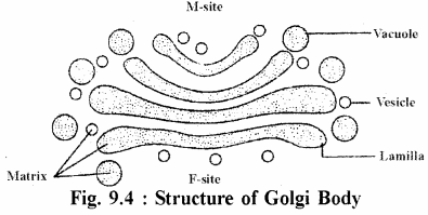
Question 6.
Give an account of structure and function of Golgi body.
Answer:
It was discovered by Cambilo Golgi (1898) in the nerve cells of owl and cat. It is also called as dictyosome or lipochondrion in plant cells and invertebrates. It is absent in prokaryotes.
Structure :
The Golgi body consists of 3 types of structures viz. –
(a) Cisternae or Lamellae – They are flat and unbranched tubes. They are found in group of 3 to 12 lamellae. They are curved. The outer surface concave and inner surface is convex. The outer concave surface is also called as trans face or maturation phase. The inner convex surface remains towards the nucleus. It is also called as cis face or maturation phase.
(b) Vacuoles : They are large & round structures and mainly situated at the ends of the cisternae.
(c) Vesicles : They are small round structures and found all over the sides of lamillae.
All the above structures are made up of single unit membrane.
function:
- It concerns with the secretory activities of the cells. It is more developed in the secretory cells like pancreatic cells, mucous cells, mammary gland cells, thyroid cells etc.
- It helps in synthesis, storage and secretion of many proteins such as albumin, enzymes, hormones, mucous etc.
- It forms the lysosomes.
- it forms cell plate during cell division.
- It forms acrosome part of the sperms.
- It forms cortical granules in the ova.
- It helps in the formation of lipoproteins.