Rajasthan Board RBSE Class 12 Biology Chapter 30 Man-Movement & Locomotion
RBSE Class 12 Biology Chapter 30 Multiple Choice Questions
Question 1.
The exoskeleton of animal is –
(a) Skull
(b) Ribs
(c) Nails
(d) Sternum
Answer:
(c) Nails
![]()
Question 2.
Matrix of a bone is of which protein –
(a) Chondrin
(b) Ossein
(c) Fibrin
(d) Retin in
Answer:
(b) Ossein
Question 3.
The function of the skeleton is –
(a) Protection of soft organs
(b) Provide space for muscle attachment
(c) Formation of blood corpuscles
(d) All of the above
Answer:
(d) All of the above
Question 4.
Reason of the pendular movement of cilia is –
(a) Sliding microtubules
(b) Contraction of microfibres
(c) Extension of cell wall
(d) Change in positions
Answer:
(b) Contraction of microfibres
Question 5.
According to sliding filament theory, the responsible molecule to decrease the length of muscle at the time of muscle contraction is –
(a) Collagen
(b) Actin
(c) Myosin
(d) Tyutin
Answer:
(b) Actin
![]()
Question 6.
Type of elbow joint is –
(a) Immovable joint
(b) Hinge joint
(c) Amphiarthrosis joint
(d) Pivot joint
Answer:
(b) Hinge joint
Question 7.
Contractile protein is –
(a) Troponin
(b) Myosin
(c) Tropomyosin
(d) All of the above
Answer:
(b) Myosin
Question 8.
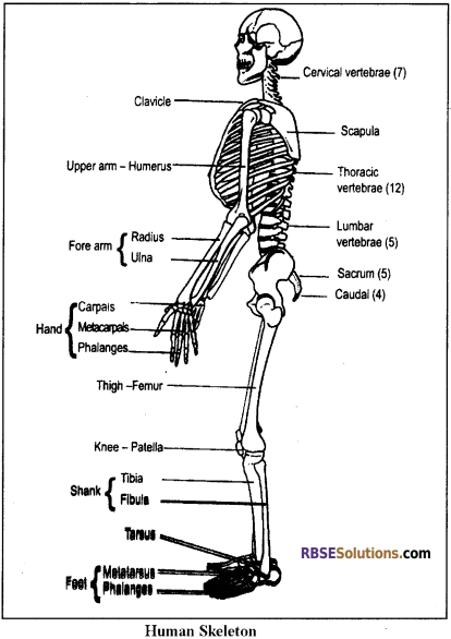
Number of bones present in the posterior limbs of man –
(a) 14
(b) 24
(c) 26
(d) 30
Answer:
(d) 30
![]()
Question 9.
Storage of which substance in an anaerobic contraction of muscles become painful.
(a) Calcium ion
(b) Myosin
(c) Lactic acid
(d) Creatine phosphate
Answer:
(c) Lactic acid
Question 10.
Presence of which ions is essential for –
(a) Calcium
(b) Sodium
(c) Ferrous
(d) Potassium
Answer:
(a) Calcium
RBSE Class 12 Biology Chapter 30 Very Short Answer Type Questions
Question 1.
Write the structural and functional unit of muscle.
Answer:
Sarcomere.
Question 2.
By which structure muscle joint with a bone?
Answer:
Tendon.
Question 3.
By which process bone joints to bone?
Answer:
Ligament.
![]()
Question 4.
How many vertebrae from sacrum bone of man?
Answer:
Five Vertebrae.
Question 5.
How many bones participate in the formation of the skull of man?
Answer:
29 bones.
Question 6.
Give names of the main substances stored in the bones.
Answer:
Calcium phosphate, calcium carbonate, magnesium phosphate.
Question 7.
Which type of energy transfer occurs in muscular function?
Answer:
Chemical energy into mechanical energy.
RBSE Class 12 Biology Chapter 30 Short Answer Type Questions
Question 1.
What are cartilaginous bones? Explain.
Answer:
The bones are of 3 types:
1. Membranous or Investing bones –
- They are formed by ossification in the membranes situated below the embryonic skin.
- They invest soft cartilaginous parts of the skeleton & provide them strength.
- Example: Flat bones of skull, clavicle, bones of digits etc.
2. Cartilaginous or Replacing bones –
- It is formed in place of hyaline cartilage.
- The matrix of the cartilage is destroyed by special osteoclasts.
- Example: Bones of limbs, vertebral column, some bones of the skull.
3. Sesamoid bones –
- It is formed by the ossification in the ligament.
- Example: Patella’s bone.
![]()
Question 2.
Write the main functions of a Skeleton.
Answer:
Functions of the Skeleton:
- The Skeleton forms a rigid framework and supports the body.
- Skeleton provides a base for attachment of muscles.
- Skeleton acts a lever during various movements of the body and also help in locomotion.
- It protects a number of delicate organs such as brain, heart, lungs and spinal cord. 5. Blood corpuscles are produced in the red bone marrow
Question 3.
Write a note on the sternum.
Answer:
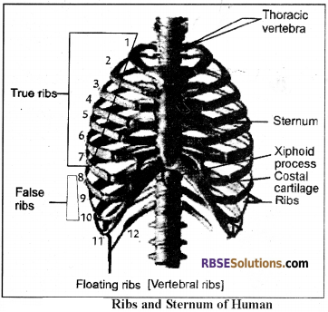
Sternum or Breast Bone:
- It forms a respiratory basket along with ribs & thoracic vertebrae.
- It is a flat bone which has distinct flat anterior & posterior sides.
- It has 3 parts –
- Manubrium – Upper part, the clavicle & ribs are attached to it.
- Sternum body or Glandiolous – Middle part, 2nd, 3rd, 4th 5th, 6th & 7th ribs get attached to it.
- Xiphoid process or Ensiform – Posterior part, small & triangular plate.
- The ribs articulate its ends.
![]()
Question 4.
Draw a neat and labelled diagram of the pelvic girdle.
Answer:
Question 5.
Differentiate between ligament and cartilage.
Answer:
| Ligament | Tendon |
| It connects bone to bone | It connects muscle to bone |
| Presence of Elastin protein | Presence of collagen Protein |
| Made up of yellow | Made up of white fibrous tissue collagen fibres |
Question 6.
How muscle gets excited about contraction?
Answer:
Excitation for Muscle Contraction:
- Nerve impulse promotes releasing of Neurotransmillar Acetylcholine the ends of the axon at Neuromuscular junction.
- This chemical changes permeability in the plasma membrane of muscle towards Na+.
- Due to this, Na+ enters into the muscle cell and causes a change in charge (axon potential), which is +ve inside the plasma membrane.
- In normal condition, there is -ve charge inside the inner surface of the plasma membrane.
- This positive change is transferred on the whole plasma membrane of muscle and generates an action potential, which is known as Excitation state of muscles.
Question 7.
If all joints of an arm of man become non-movable than what happens?
Answer:
Limb movement & Locomotion will become impossible.
Question 8.
What is osteoporosis?
Answer:
Osteoporosis:
Main characteristics of the disease are the loss of bone mass means deficiency of organic part (matrix) and minerals (calcium) in the bones. The bone becomes thin, weak and loses elasticity and strength. Chances of bone fracture are increased and slight jark or impact may lead to bone fracture.
It affects the whole body skeleton but pelvic, wrist, vertebrae etc are most adversely affected parts. It increases with the age but deficiency of estrogen also caused osteoporosis in old women. It may also be caused by hormones like calcitonin, PTH and glucocorticoids.
![]()
Question 9.
What is the source of energy for muscle contraction?
Answer:
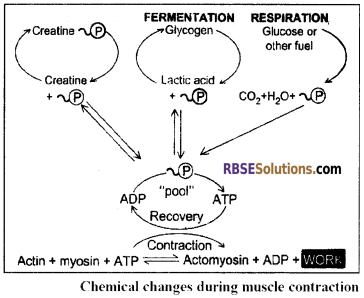
Energy Sources for Contraction:
- Both muscle contraction & relaxation require energy and the main source of energy is ATP. The muscles have less number of stored ATP. Hence, during contraction ATP is obtained from other sources.
- The creatine – phosphate found in the muscles provides energy instantly because it has one high energy bond. It is also called as phosphagen. This process is catalysed by an enzyme creatine kinase.
- The energy required in muscle contraction is obtained by oxidation of glucose but due to deficiency of O2, this source fails to provide energy for a long period. Hence, the fermentation of the glycogen is the long-lasting source of the energy for muscle contraction.
- Glycogen fermentation results in the formation of lactic acid. This lactic acid is transported into the liver by the blood. In the liver, 80% lactic acid is recovered into the glycogen (Glyconeogenesis) and the remaining 20% lactic acid is oxidized into CO2 & H2 in which energy is released. This energy is used in the glyconeogenesis.
Question 10.
If nerve going to skeletal muscle is cut, what will be an effect on contraction?
Answer:
There will be no contraction.
RBSE Class 12 Biology Chapter 30 Essay Type Questions
Question 1.
Write the structure of skeletal muscle in detail.
Answer:
Structure Of Skeletal Muscle:
- Each skeletal muscle is spindle-shaped & has 2 distinct ends viz., Origin & Insertion. The origin & insertion remain attached to the bones with the help of tendon (cord-like structure) or aponeuroses (membrane-like structure).
- The skeletal muscles of the tongue & upper part of the oesophagus are not attached to the skeleton. These muscles are also called as fatigued muscles or phasic muscles or striated muscles or somatic muscles.
- Their structural unit is called as muscle fibre.
- The muscle fibre is multinucleated or syncytial & bounded by sarcolemma. The muscle fibres are found in group & each group is called as fasciculus. The fasciculus is bounded by endomysium.
- The fasciculi are also found in the group and each group of fasciculi is bounded by perimysium which is made up of connective tissues.
- The whole skeletal muscle is bounded by an epimysium.
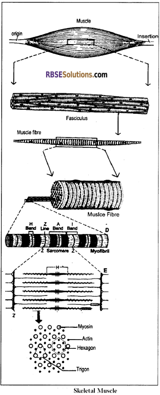
Fine structure of muscle Fibre:
- The maximum length of the muscle fibre is 30 cm (1 – 30 cm) and the diameter is 0.01 to 0.1 mm. It is bounded by a sarcolemma or plasmalemma or myolemma. Its Sarcoplasm has many mitochondria which are called as acrosomes. The ER found in the sarcoplasm is called as Sarcotubules.
- The sarcoplasm has three soluble proteins viz., myogenin, myoglobin & my albumin.
- The sarcoplasm stores glycogen. In addition, it has less quantity of Na, Ca, P& Mg & more quantity of K.
- The muscle fibre has many myofibrils. The length of the myofibril is similar to the muscle fibre but its diameter is 1 to 3µ.
- The Myofibril has alternate A – bands (Anisotropic) & I – bands (Isotropic).
- There is a Z – line (zigzag) in the middle of the I – band which is also called as Krau’s membrane.
- The middle zone of the A-band is called an H – zone which has a central M – line.
- The distance between two Z – lines is called sarcomere which is a functional unit of the muscle.
- Each sarcomere includes one complete A – band and 2 halves l – bands on both the sides (12 + A + I/2).
- The A – band is made up of mainly myosin filaments. The length of the myosin filament is 1.5µ & the diameter is 100Å.
- The l-band is made up of mainly actin filaments. The length of the actin filament is 2µ & the diameter 50Å.
0.2µ of both the ends of the actin filaments size remain in the A – bands. - During the resting phase, the length of the A-band is 1.5µ & of the half I-band is 0.8µ. Hence, the length of the sarcomere is 3.1µ.
- In the transverse section of the myofibril, through the end of A-band, has 5000 filaments (Both actin & myosin).
- Each myosin filament is surrounded by 6 actin filaments (Hexagon). Similarly, one actin filament is surrounded by 3 myosin filaments (Trigon) The myosin filaments are provided with cross-links or bridges.
- The cross-links remain directed towards the actin. They are attached at an angle of 60° & are equally spaced by 60 – 70 Å. The H – zone is without cross-links.
![]()
Question 2.
What is the joint? Describe various kinds of joints found in man.
Answer:
Joint:
- In the vertebrates, joints are the places where two bones articulate with each other or bone and cartilage articulate with each other. In other words, the contact place between two or more bones or between bone and cartilage is known as Joint.
- These joints facilitate movements of the body parts.
On the basis of movements, the Joints are of three types –
Synarthrosis or fixed or Immovable Joints:
- The articulating bones are held together by a dense bundle of tough and white fibrous connective tissue which cannot be stretched or extended.
- No movement is possible and hence called as fixed joints or synarthrosis.
- Examples: Joints between the bones of the skull which are called Sutures.
Amphiarthrosis or partially movable joints:
- It is a tough joint in which bones are joined by a disc of white fibrous cartilage which can be stretched a little.
- Limited movement is possible.
- Example: Pubic Symphoisis
- It is of two types –
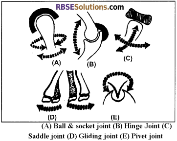
- Pivot Joint – Joints between Atlas and axis vertebrae. It provides lateral movement.
- Gliding Joint – In such joint, end of one bone glides over a certain portion of another bone. Movement in different directions is found. Joints between tarsal bones of the ankle, carpal bones in the wrist and in between sternum and clavicle.
Synovial or Diarthrosis ou movable Joints:
- The Bones joined by synovial joints can move in one or more directions freely. A highly smooth thin layer of hyaline cartilage is found on the surface of the bone where joint occurs.
- This cartilage minimizes the friction between the bones. Space is found between the joints of bones, which is called the synovial cavity. Bones are joined with each other by many ligaments. Ligaments fuse together and form a capsule which is covered by synovial membrane.
- It secretes mucin containing synovial fluid. This fluid provides nutrition to hyaline cartilage and lubrication to joints.
- It is of three types –
- Ball and Socket Joint – It this joint ball-like the end of one bone fits into the socket of another bone. Movement of bone with the ball head in various planes is possible. Example: Shoulder Joint and Hip Joint.
- Hinge joint – Resemble the hinges of doors. Movement in one direction only. Examples – Elbow joint, knee joint and finger joint, occipital condyle and atlas joint.
- Ellipsoidal joint – Movement of one bone over another bone, allowing movements in around two axes. Joints between metacarpals and phalanges in hand, and metatarsals and phalanges in the foot. Another example is the joint between radius and carpals.
![]()
Question 3.
Describe the mechanism of muscle contraction with diagrams.
Answer:
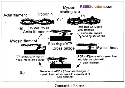
Mechanism of Muscle Contraction:
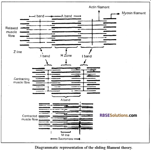
Sliding Filament Theory:
- The process of muscle contraction can be explained with the help of “Sliding filament theory” which was given by A.F. Huxley, J. Hensen & H.E. Huxley.
- According to it, the myosin filaments remain stationary & the actin filaments slide over the myosin with the help of cross-links.
- During contraction, the length of the A – band remains unchanged but the length of the I – band & H – zone get reduced.
- In H – zone, the actin filaments slide over each other.
- The contraction results in the shortening of the sarcomere.
- The relaxation of the muscle involves reverse sliding of the actin filaments to obtain an original position.
Physiology of Muscle Contraction:
The process of muscle contraction & Relaxation involves four phases.
1. Excitation:
- Nerve impulse promotes releasing of Neurotransmillar Acetylcholine the ends of the axon at Neuromuscular junction.
- This chemical changes permeability in the plasma membrane of muscle towards Na+. Due to this, Na+ enters into the muscle cell and causes a change in charge (axon potential), which is +ve inside the plasma membrane. In normal condition, there is -ve charge inside the inner surface of the plasma membrane.
- This positive change is transferred on the whole plasma membrane of muscle and generates an action potential, which is known as Excitation state of muscles.
2. Excitation – Contraction Coupling:
- This is the process in which action potential promotes contraction in a muscle cell. This action potential instantly transfers to the T – system.
- Due to this potential, Ca++ released from the sarcoplasmic reticulum and join with troponin C.
- It results in configurational changes in Troponin atoms.
- As a result, tropomyosin and troponin, both are released from the active site of actin.
- As soon as active site is made free the myosin filament joins with actin and contraction process starts.
3. Contraction:
- Contraction occurs by sliding filament process, which is initiated by joining of actin & myosin transverse bridge.
Just before joining of active filament at the active site, one atom of ATP joins at the head of the transverse bridge. ATP present in myosin head breaks down into ADP+ Pi by ATPase.
ADP+ Pi remain attached or joined with the head after that myosin head joins with the active site of the actin filament. - There are conformational changes in the head due to joining of head and active site. Due to this, there is bending in the head (just as we bend our finger) and actin filament is pulled towards the centre of the sarcomere (H zone) and energy is obtained to do this by breaking down of ATP.
- Due to bending of head, ADP+ Pi are also released and as they are released, instantly new ATP joins with the head.
- Due to joining of the head with ATP, it separates from actin. Again ATP is broken down Myosin head now joins again at the new active site and this process is repeated.
- Due to repeated acts of the head of the transverse bridge, active filament slides and leads to contraction. In contraction many filaments. Cross bridges (transverse bridges) participate. The contraction in muscle tissue (filament) continues till Ca++ remain attached with troponin.
4. Relaxation:
- When contraction through nerve impulse is stoped in muscle then Ca++ is sent back to muscle sarcoplasmic reticulum, which results in troponin – C free from Ca++ and active sites of actin filament are blocked.
- Now there is no joining or bonding of actin and myosin filament. Filaments come into their original position and muscles relax.
![]()
Question 4.
Write the structure of girdles and their significance.
Answer:
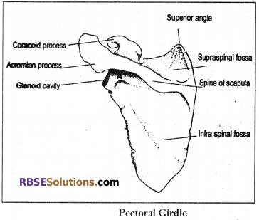
Pectoral Girdle:
- It supports fore limbs & protects chest organs.
- It has two equal halves.
- Each half has a clavicle & a scapula-coracoid bones.
- The clavicle is a rod-like bone which is also called as collar bone.
- The scapula is a broad & flat bone which is found in all mammals.
- The coracoid is get fused with the scapula & found in the form of a coracoid process.
- The supra scapula is absent in the pectoral girdle of Man.
- The dorsal side of the scapula has a spine. The anterior & ventral extension of the spine is called an acromial process.
- The posterior process of acromial in the rabbit is called the acromion process.
- The head of the scapula-coracoid has a cavity, the glenoid cavity.
- The head of the humerus bone articulates to the glenoid cavity.
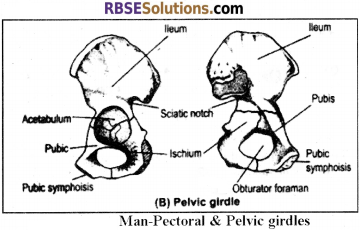
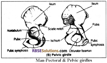
Pelvic Girdle:
- It supports hind limbs.
- It has 2 equal halves & each half is called as os-innominate.
- Both the os-innominate remain united by pubic symphysis which is made up of fibrous cartilage.
- The pelvic girdle of the rabbit is W – shaped.
- Each os – innominate of the rabbit has 4 bones – Ilium, Ischium, Pubic & Cotyloid.
- Each os – innominate has a groove, the acetabulum. The head of the femur articulates with it.
- In the rabbit, the pubic bone does not participate in the formation of the acetabulum. There is a small cotyloid bone between the acetabulum & the pubic.
- In man, cotyloid bone is absent. Hence, each os-innominate has 3 bones – Ileum, Ischium & Pubic. All of them participate in the formation of the acetabulum.
- There is an obturator foramen between pubic & ischium bones.