Rajasthan Board RBSE Class 12 Biology Chapter 42 Bio-Medical Technologies
RBSE Class 12 Biology Chapter 42 Multiple Choice Questions
Question 1.
Which of the following is used to measure blood haemoglobin?
(a) Haemocytometer
(b) Haemoglobinometer
(c) Westergren method
(d) Wintrobe method
Answer:
(b) Haemoglobinometer
![]()
Question 2.
In which of the following disease, number of leucocytes increases?
(a) Tuberculosis
(b) Typhoid
(c) Measles
(d) Blood cancer
Answer:
(d) Blood cancer
Question 3.
Which is used to diagnose the heart-related diseases?
(a) EEG
(b) ECG
(c) RIA
(d) CAT scan
Answer:
(b) ECG
Question 4.
Which of the following radiations are used in a CT Scan?
(a) α – rays
(b) β – rays
(c) γ – rays
(d) X – rays
Answer:
(d) X – rays
![]()
Question 5.
Full name of MRI is:
(a) Multiple Resonance Imaging
(b) Magnetic Radio Imaging
(c) Magnetic Resonance Indicator
(d) Multiple Radio Imaging
Answer:
(c) Magnetic Resonance Indicator
RBSE Class 12 Biology Chapter 42 Very Short Answer Questions
Question 1.
What is used for Total leucocyte count?
Answer:
Neubauer’s haemocytometer.
Question 2.
What is Leucocytosis?
Answer:
When the number of WBC becomes more than normal.
Question 3.
In which diseases, the value of ESR increases.
Answer:
Rheumatoid Arthritis, Multiple Myeloma, TB etc.
![]()
Question 4.
Name the instrument that records heartbeats.
Answer:
Stethoscope.
Question 5.
EEG is related to the diagnosis of which organ functioning?’
Answer:
Brain.
Question 6.
Which is used in place of X – rays in MRI.
Answer:
Magnetic Resonance:
Imaging uses the electric charge and low magnetic field of the nuclei of the hydrogen atom generated in an environment of a very strong magnetic field and radio waves. Hydrogen atoms are present in proteins and water molecules.
Question 7.
Which crystals are used in sonography?
Answer:
Lead Zirconate Crystals.
![]()
Question 8.
Which functions as labelling molecule in RIA?
Answer:
Radioisotopes substance.
RBSE Class 12 Biology Chapter 42 Short Answer Questions
Question 1.
Explain in short about the Westergren method of ESR.
Answer:
Erythrocyte Sedimentation Rate (ESR):
- If a blood sample mixed with an anticoagulant like Trisodium citrate and kept undisturbed for some time in an ESR measuring tube, blood cells being denser than plasma, moves towards the bottom of tubes and settler down. The rate of setting down of the blood cells is called Erythrocyte Sedimentation Rate. The ESR measurement can be done by two methods.
- Westergren method
- Wintrobe method
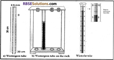
- In most of the pathological laboratories, only the Westergren method is used for ESR testing. Hence, here this method is described.
- Westergreen tube is filled up to zero points with the blood containing anticoagulants. This tube is kept legitimately in a verticle position in ESR stand.
- After an hour the upper level of erythrocytes is read in the ESR tube. This is the value of Erythrocyte sedimentation rate.
- The ESR value of a healthy person is as follows:
- Male = 0 – 16 mm per hour.
- Female = 0 – 20 mm per hour.
- If the rate of ESR is more than the normal value, it indicates towards irregularities in the body. Value of ESR increases in many chronic diseases like tuberculosis and inflammatory diseases like Rheumatoid arthritis, multiple myeloma or allergy, lymphatic inflammation etc.
- Moreover, the value of ESR increases during pregnancy, Anaemia and with an increase in Age. These days Automated mini ESR machine is used to enhance the accuracy in the value of ESR. The machine works at a controlled temperature of 18°C. In the traditional methods, temperature regulation is not possible and a chance of 25 – 30% error exists.
![]()
Question 2.
Write the medical significance of Differential Leucocyte Counts.
Answer:
Medical Significance:
- Any irregularity observed in the differential counting of the leucocytes is indicative of specific disease; further specialized testing can give a proper diagnosis of the disease and thus helps in proper treatment.
Some of these conditions are described below.
| Nature of irregularity in differential leucocyte counting | Symptoms of possible diseases |
| Increase in the number of Neutrophils | Infection leading to normal pus, Sign of inflammation & dermatitis. |
| Increase in Eosinophils | Hypersensitivity or Allergy and signs of Parasitic infection |
| Increase in Basophils | Chickenpox disease |
| Increase in Lymphocytes | Whooping cough |
| Increase in Monocytes | Signs of Tuberculosis |
| The decrease in T4 Lymphocytes | Signs of AIDS |
(In General differential leucocyte count do not include T4 lymphocytes).
Question 3.
Write the use of ECG.
Answer:
Use of ECG:
- It is used to detect cardiac disorders like arrhythmias, conduction disturbances; myocardial ischemia etc.
- It reveals other findings related to life-threatening metabolic disturbances like hyperkalemia or increased, susceptibility to sudden cardiac death (Example: QT prolongation syndrome).
- It is used to study the rate of heartbeat, and cardiac disorders such as myocardial infarction (MI), coronary artery diseases etc.
Question 4.
Write the significance of Sonography technique.
Answer:
It is used to assess foetal growth and also the abnormalities in an adult body. It can provide a picture of blood flow through the beating heart.
![]()
Question 5.
Explain the role of ESR in disease diagnosis.
Answer:
Value of ESR increases in many chronic diseases such as TB, Arthritis, Allergy etc. ESR value also increases during pregnancy, Anaemia and old age.
Question 6.
Why MRI technique is better and safe than a CT scan.
Answer:
MRI provides high contrast images from all body axis. This technique can clearly differentiate between the white and grey matter of the brain.
RBSE Class 12 Biology Chapter 42 Essay Type Questions
Question 1.
Explain in detail about the process of haemoglobin measurement in blood.
Answer:
Estimation of Haemoglobin in blood:
- Estimation of haemoglobin in the blood is called Haemoglobinometry. Haemoglobin is a respiratory pigment found in red blood corpuscles. Chemically, it is a chemoprotection which is used for the transport of O2 and CO2.
- Working efficiency of a person has affected adversely if due to some reason his/her haemoglobin content drops below the normal value. A value of haemoglobin less than the normal value is an indicator of Anaemia.
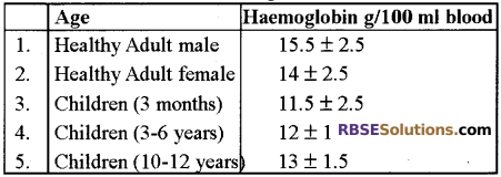
- The normal value of Haemoglobin is as follows:
| Age | Haemoglobin g/100 ml blood |
| Healthy Adult male | 15.5 ± 2.5 |
| Healthy Adult female | 14 ± 2.5 |
| Children (3 months) | 11.5 ± 2.5 |
| Children (3 – 6 years) | 12 ± 1 |
| Children (10 – 12 years) | 3 ± 1.5 |
- Haemoglobin content is measured with Haemoglobinometer. Traditionally Sahli’s Haemoglobinometer is used for this purpose.
- Photohaemoglobinometer and Autoanalyzer are also used.
- Sahli’s Haemoglobinometer:
- This device contains a graduated tube and two standard matching tubes placed in a stand. Between the two standard tubes is placed a measuring graduated tube. In the measuring tube, N/10 HCl is filled up to zero (2 g%) point.
- Now using the haemoglobin pipette 20 Jul (0.02 ml) blood is transferred to the measuring tube. Blood is thoroughly mixed with N/10 HCl already present in the tube.
- The N/10 HCl converts haemoglobin into a brown coloured substance called haematin. The measuring tube is now kept in between in the two standard tubes.
- Distilled water is added drop by drop mixing with continuous mixing to the measuring tube until the colour of the haematin solution matches with the colour of the standard tubes/comparison tubes.
- When the colour matching between the tube is ensured, haemoglobin content is measured by observing the reading on measuring graduated tube.
![]()
Question 2.
Explain the diagram of ECG and write its uses.
Answer:
Salient Features of ECG:
- The ECG is recorded on special graph paper which is divided into 1 mm2 gride like boxes.
- The normal ECG includes P wave, QRS complex and ST – T – U complex.
- The P wave is a small upward deflection and it represents atrial depolarization.
- The QRS Complex represents rapid ventricular depolarisation (systole). It includes a small downward deflection, a rapid upright stroke and a small downward deflection.
- The ST – T – U complex includes ST regiment, T wave & U wave. It represents the ventricular repolarisation (diastole).
- The J point is the junction between the end of the QRS complex and the beginning of ST-segment.
- Trial repolarization is too low in amplitude to be detected.
- There are four major ECG Intervals –
- R – R interval: It is used to compute the heartbeats per minute.
- P – R Interval: It is used to measure the time between atrial & ventricular depolarisation.
- QRS interval: It reflects the duration of ventricular depolarisation & atrial repolarisation.
- QT interval: It includes both ventricular depolarisation and repolarisation times.
- The QRS Complex is subdivided into specific deflections or waves.
- The initial negative deflection is termed as Q – wave.
- The first positive deflection is termed as R – wave A negative deflection after the R – wave is termed as S – wave.
- An entirely negative QRS complex is termed as QS wave.
- Normal T wave is a dome-shaped upward deflection.
- Normal U wave is a small round upward deflection that follows the T – wave.
Question 3.
Write an explanatory note on MRI.
Answer:
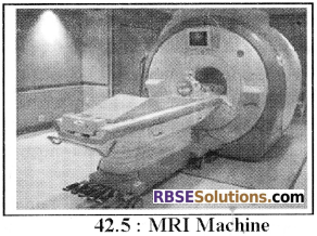
Magnetic Resonance Imaging (MRI):
- MRI was discovered by F. Bloch and E.M. Purcell independently and they shared the Nobel prize in 1952.
- In this technique, there dimensional images of body organs are obtained without using X – rays & other radiations.
- MRI detects water because it focuses on the behaviour of hydrogen atoms in water molecules.
- MRI distinguishes between water-poor & water abundant tissues. The tissues with little water such as bones & teeth do not appear in MRI.
- This technique is based on the natural behaviour of protons of hydrogen atoms and the most abundant source of protons are the hydrogen atoms in the water molecule.
- MRI is used mainly to study organs like brain & spinal cord, to examine joint injuries & slip discs and to visualize minute cancerous tumours.
- MRI is based on the phenomenon known as nuclear magnetic resonance (NMR).
- During MRI testing, the patient is positioned in a two-meter wide chamber of an MRI scanner.
- This chamber is surrounded by a large cylindrical electromagnetic field that produces a very strong magnetic field and waves.
- Because of the very high magnetic field, nuclei of the hydrogen atom get activated and release radio signals.
- These signals are processed by a computer. Through the processing of radio signals, thin high contrast images are obtained.
- The MRI images are much better than the images obtained by CT scan.
- They provide high contrast, using MRI technique, images from all body axis can be obtained.
- Although MRI is a costly technique it is very helpful for the diagnosis and study of the brain and spinal cord.
- This technique can clearly differentiate between the white and grey matter of the brain.
![]()
Question 4.
What is RIA? Explain its procedure and uses.
Answer:
Radio Immuno Assay (RIA):
- Radioimmunoassay is an analytic technique which is used from the last many years.
- Here, the molecule which is to be analysed, which acts as an antigen is marked with a radiolabelled substance.
- The labelled and normal antigen molecules are allowed to react with a specific antibody.
- A comparative analysis of the reaction is done. In this technique, the radioisotope is used as a marker molecule.
- This technique was invented by Rosalyn and Yalow. Now – a – days it has become an important diagnostic technique.
- This is especially important for the analysis of those biochemical factors that are present in very minute concentration (microgram, nanogram or picogram) and cannot be analysed by the traditional gravimetric and volumetric processes.
- The radioisotopes used in radioimmunoassay are high specificity molecules, which provide very high sensitivity to this technique.
- In this technique, the standard solution of different concentration of normal molecules of the substance to be analysed is used with the solutions of the radiolabelled substance of similar concentrations.
- This mixture is allowed to react with antibodies. At the stage of equilibrium, antigen-antibody complexes are absorbed by the suitable reagent.
- The precipitated and supernatant parts are separated and by measuring the radioactivity, the concentration of the substance is estimated.
- A major characteristic of Radio Immuno Analysis is that the patient does not suffer from any side effect.
- He is never treated with the radioisotopic molecule because the whole process is executed outside the body.
Uses of RIA:
- Using this technique, the concentration of important biological components like vitamins (B12, Folic acid), hormones (Thyroxine, Triodothyroxine, T3, Cortisol, Testosterone, Estrogen, Tropic hormone etc.) drugs (Digoxin, Digitoxin etc.) and
antigenic substances can be determined. - For the diagnosis of abnormalities of endocrine glands, radioimmunoassays are very useful. For example presence of an excessive amount of some hormones in the blood may be a result of hyperactivity of endocrine gland or due to the impact of the tropic hormone, such questions can only be answered by this technique.
- This technique is useful for the diagnosis of insulinoma, tumour etc which will help in the proper treatment of these diseases.