RBSE Solutions for Class 9 Science Chapter 6 Structure of Living Organisms are part of RBSE Solutions for Class 9 Science. Here we have given Rajasthan Board RBSE Class 9 Science Chapter 6 Structure of Living Organisms.
| Board | RBSE |
| Textbook | SIERT, Rajasthan |
| Class | Class 9 |
| Subject | Science |
| Chapter | Chapter 6 |
| Chapter Name | Structure of Living Organisms |
| Number of Questions Solved | 81 |
| Category | RBSE Solutions |
Rajasthan Board RBSE Class 9 Science Chapter 6 Structure of Living Organisms
Structure of Living Organisms Textbook Questions Solved
Objective Type Questions
Question 1.
Which organelle of the cell is known as Suicidal Bag?
(A) Mitochondria
(B) Lysosome
(C) Ribosome
(D) Golgi body
Answer: (B)
Question 2.
Which organelle of the cell is known as the powerhouse of the cell?
(A) Mitochondria
(B) Lysosome
(C) Ribosome
(D) Nucleus
Answer: (A)
Question 3.
Which scientist discovered the nucleus?
(A) Robert Brown
(B) Robert Hook
(C) Leuwen hock
(D) Schliden
Answer: (A)
Question 4.
In which phase of cell cycle, DNA is synthesised:
(A) G – I phase
(B) S phase
(C) M phase
(D) G – II phase
Answer: (B)
Question 5.
Tissue which gives flexible strength to plants is:
(A) Parenchyma
(B) Collenchyma
(C) Sclerenchyma
(D) None of the above
Answer: (B)
Structure of Living Organisms Very Short Answer Type Questions
Question 6.
Name the scientist who saw the living cell, for the first time?
Answer
Leeuwenhoek saw the living cell for the first time.
Question 7.
Name any two unicellular organisms?
Answer
Two unicellular organisms are:
(a) Amoeba
(b) Chlamydomonas
Question 8.
Name the largest cell of the human body?
Answer
The largest cell of the human body is Neuron (nerve cell)
Question 9.
What is the function of the cell wall in a plant cell?
Answer
(a) Cell wall provides rigidity, structural strength and definite shape to the cell.
(b) It helps in transport of various substances across it.
Question 10.
On the basis of pigments, how many types of plastids are present in plants?
Answer
On the basis of pigments, three types of plastids are present in plants.
They are:
- Leucoplasts (Colourless plastids)
- Chloroplasts (green plastids)
- Chromoplasts (Coloured plastids, other than green)
Question 11.
What is the function of the ribosome in a cell?
Answer
The function of ribosomes is to synthesise proteins in the cell. Both the bounded and free ribosomes are involved in this synthesis.
Question 12.
What type of division takes place in somatic cells?
Answer
In somatic cells, mitosis-cell division takes place.
Question 13.
Why meiosis division is known as a reductional division?
Answer
In meiosis, cell division the first division reduces the chromosomal number to half and is, therefore, called the reductional division. It produces two haploid cells from the parent diploid cell.
Question 14.
In plants during cell division, cytoplasm is divided in which phase?
Answer
The division of cytoplasm occurs after karyokinesis in the cytokinesis phase of mitosis.
Question 15.
Which type of substance is deposited on the cell wall of collenchyma tissue?
Answer
Cellulose and Pectin are deposited on the cell wall of collenchyma tissue.
Structure of Living Organisms Short Answer Type Questions
Question 16.
What are unicellular and multicellular organisms? Give example.
Answer
A unicellular organism has a single cell carrying out all life process. This is unable to exhibit a wide range of different functions and easily susceptible to damage, that can lead to the death of the organism.
For example: Amoeba, Chlamydomonas etc.
A multicellular organism has different cells carrying out different functions. They are more efficient and carries out no. of activities. They have a greater capacity of survival and they are continuously replaced.
For example: Cat, Dot, Human being etc.
Question 17.
Explain the cell theory?
Answer
Cell theory: A cell is the smallest structural and functional unit of any living organism. The concept that the cell is the basic unit of life is called the cell theory. At first, German Scientist, M.J.Schleiden and Theodor Schwann established cell theory. The cell theory was further expanded by Virchow, by expressing that all cells arise from pre-existing cells.
Thus the modern version of the cell theory summarized as follows:
- All living things, whether plants or animals are composed of cells and their products.
- A cell is the smallest unit of structure and function, which arise from pre-existing cells and their continuity is maintained through the genetic material.
- All cells are basically alike, found after chemical analysis and studying the functions of different components.
Following are the concepts of the modern cell theory:
- The body of each living being is composed of one or more cells and cell products.
- The cell is the fundamental unit of all biochemical reactions.
- A cell is also a hereditary unit because it contains genetic material in it.
- New cells are formed from the division of pre-existing cells.
- All organisms start life from a single cell.
- All organisms function through the activities of cells.
Question 18.
Explain the structure and function of Mitochondria?
Answer
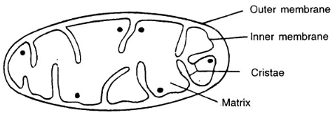
Mitochondria: They are small bodies that occur in large numbers.
A mitochondrion consists of:
- an outer membrane
- an inner membrane
- inner membrane folded inwards to form the cristae.
- The cristae lie in a ground substance called as a matrix.
Functions of Mitochondria:
- It generates energy for various activities of the cell.
- Whenever the cell requires energy, ATP molecule breaks down generating energy to be used by the body, in order to make chemical compounds and perform mechanical work.
- Mitochondria have their own DNA and ribosomes hence, are able to make some of their own proteins.
Question 19.
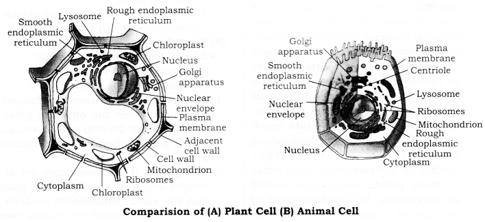
Write the four difference between Animal and Plant cell?
| Plant Cell | Animal Cell |
| 1. Cells are comparatively larger in size. | 1. Cells usually smaller in size. |
| 2. Generally rectangular in shape. | 2. Generally oval in shape. |
| 3. Cell wall made of cellulose is present. | 3. The cell wall is absent. |
| 4. Plastids are present in the cytoplasm. | 4. Plastids are absent. |
| 5. Vacuoles-generally only one, large vacuole is present. | 5. Vacuoles-generally absent, if present, they are more in number and smaller. |
Question 20.
Why lysosomes are known as suicide bags?
Answer
Lysosomes are tiny vesicles, each of which is surrounded by a single membrane and contains enzymes, which are capable of breaking down all sort of organic materials. They are called the ‘suicide bags’ of the cell, as they contain hydrolytic enzymes which can digest incoming food materials; remove the foreign bodies, toxic molecules, and debris; breakdown wore out cells and cell organelles to component molecules, for building the new organelles and cells.
Question 21.
Describe the structure and function of the nucleus?
Answer
Robert Brown in 1831 discovered the nucleus in the cell.
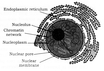
Structure: The nucleus is of large size, more dense, spherical or oval and prominent organelle of the cell.
The structure of the nucleus can be divided into three parts:
1. Nuclear membrane: Nucleus is enclosed by a double-layered nuclear membrane. In bacteria and blue-green algae, the nuclear membrane is not found.
2. Nucleoplasm: In nucleus is present a thick jelly-like semi-fluid nucleoplasm. In nucleus network of fine thread-like structures called chromatin network. At the time of cell division, the chromatin takes on the form of thread-like structures called chromosomes. The number of chromosomes is fixed for every species of animals and plants. The man has 23 pairs of chromosomes. Wheat has 21 pairs of chromosomes. The two partners of chromosomes pair are called homologous chromosomes.
-
- Chromosomes: Chromosomes are composed of specific proteins and a nucleic, acid called Deoxyribose, Nucleic Acid (DNA) Genes are segments of DNA. Genes control and carry the hereditary characters.
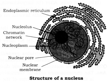
3. Nucleolus: Nucleolus contains RNA, commonly there is a single nucleolus, but two or more may be present. The nucleolus is the seat for the synthesis of cytoplasmic, Ribosomes. During cell, division nucleolus disappears in early stages and reappears on the same location in the daughter nuclei.
The functions of the nucleus are as follows:
- The nucleus controls the activities of the cell, through its DNA molecule.
- The main function of the nuclear reticulum is participation in cell-division.
- Genes are located in a linear fashion in chromosomes. The genes are composed of DNA. Genes control and carry the hereditary characters from one generation to another generation.
- Nucleolus assists in protein synthesis. Formation of ribosomes takes place in the nucleolus.
Question 22.
Explain the Cell Cycle?
Answer
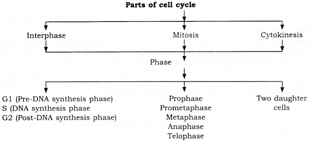
Cell Cycle: It is a sequence of events which occur between the formation of the cell and its division into the daughter cell.
Question 23.
Explain the method of cytokinesis in plant and animal cell?
Answer
The division of the cytoplasm occurs after the Karyokinesis, and differs in animal cells and plant cells:
- In animal cells: Towards the end of telophase, a peripheral furrow appears in the cytoplasm, precisely in the middle of the cell. The furrow deepens, and gradually reaches the centre, dividing the cell into two daughter cells.
- In plant cells: After the karyokinesis, small membrane-bound vesicles settle in the form of a row in the middle of the cytoplasm. The vesicles fuse to form a cell plate. The latter changes to middle lamella on either side, of which is the deposited material for the cell wall.
The formation of cell wall divides the cell into two:
Plant Cell:
- No centriole present
- No aster forms
- Cell plate forms
- No furrowing of cytoplasm at cytokinesis
- Occurs mainly at meristems.
Animal Cell:
- Centrioles present
- Asters form
- No cell plate forms
- Furrowing of cytoplasm at cytokinesis,
- Occurs in tissues, throughout the body.
Question 24.
Explain the metaphase of the mitotic division with a diagram?
Answer
The chromosomes arrange themselves at the equatorial or metaphase plate. This is the fluid area in the centre of the cell.
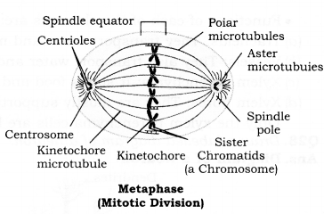
The orientation of the chromosomes is such that their centromeres lie along the equator, while the chromatids extend freely in any direction in the surrounding cytoplasm. This is called astral mitosis. In animal cells and cells of the lower plants, centrioles and asters are present. Such mitosis is called astral mitosis or amphiastral mitosis.
In cells of higher plants, however, centrioles and asters are absent, hence, the mitosis is called anastral mitosis. Metaphase is considerably shorter than prophase, but slightly longer than anaphase.
Question 25.
Explain the Anaphasic Movement in cell division?
Answer
Anaphasic movement: In Anaphase, centromere splits into two as a result of which each chromosome separates into two sister chromatids. These sister chromatids or daughter chromosomes move towards the opposite poles. This movement is due to repulsion between the centromeres and contraction of the spindle fibres. This movement is known as Anaphasic movement.
Question 26.
Write the significance of Meiosis?
Answer
The significance of Meiosis.
- Due to this division, the gametes formed are haploid. Hence, meiosis is an important and essential part of the life history of living beings.
- During fertilisation, the gametes on fusion form diploid zygote. Thus the original number of chromosomes in somatic cells is restored.
- New characters are introduced in the new generation due to crossing over of the genes.
- Due to changes in parental characters, variations occur, which are necessary for the evolution process.
Question 27.
Explain the structure and functions of Xyrem?
Answer
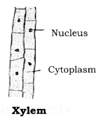
Xylem is a complex permanent tissue which conducts water and mineral salts upward from roots to leaves.
Structural elements of xylem:
- Tracheids are elongated, dead cells with large cavities and possess highly lignified, thick walls. Depending upon the kind of thickening, tracheids may be annular, spiral, reticulate, scalariform and pitted.
- Vessels are broader as compared to the tracheids. These are composed of many cells, joined end to end with their walls perforated to give it a tube-like appearance. They also show thickening like the tracheids.
- Xylem Fibres (= Wood fibres): They represent selerenchyma fibres present in xylem. The fibres are mechanical in function.
- Xylem Parenchyma (= Wood parenchyma): It is parenchyma present in xylem. The cells are, however smaller and thick walled. Xylem parenchyma stores food and helps in lateral conduction of sap or water
Functions of each of the elements are:
- Tracheids: They transport water and minerals vertically.
- Vessels: They also transport water and minerals vertically.
- Xylem parenchyma: It stores food and helps in sideways conduction of water.
- Xylem fibres: They are mostly supportive in function.
Only the xylem parenchyma cells are living cells.
Question 28.
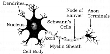
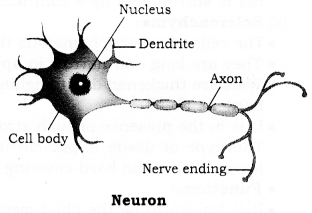
Draw a labelled diagram of a neuron.
Answer
Diagram of a Neuron:
Question 29.
Describe the various muscles found in Animals?
Answer
These consist of elongated cells called muscle fibres. It is responsible for movement in our body. It contains a special type of protein called contractile proteins, which causes movement of muscles by contracting and relaxing.
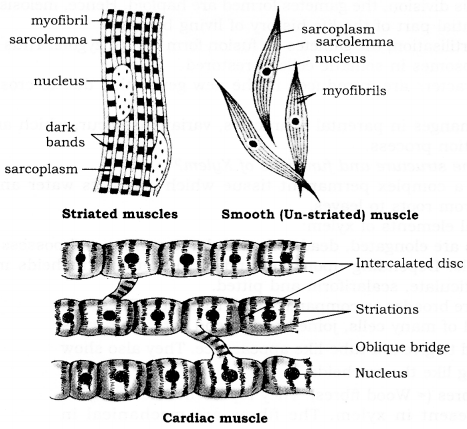
Different types of muscular tissues are given below:
(a) Striated muscle: It is also called as voluntary muscle, as we can move them by conscious will. It is mostly attached to bones and helps in body movement e.g. muscles of limbs.
- The cells are long, cylindrical, unbranched and multinucleate.
- Under the microscope, striated muscles show alternate light and dark bands or striations when stained appropriately. Hence, they are also called striated muscles.
(b) Smooth muscle or Unstriated muscle: It is also called involuntary muscle, as we cannot move them according to our will e.g. movement of food in the alimentary canal, contraction and relaxation of blood vessels, iris of the eye, in ureters and in bronchi of lungs.
- The cells of smooth muscles are long with a pointed end, uninucleate.
- These muscles do not show any dark or light band. Hence, they are also called unstriated muscles.
(c) Cardiac muscle: These are involuntary muscle present only in our heart. It performs rhythmic contraction and relaxation throughout life. The cells of cardiac muscles are cylindrical, uninucleate and branched.
Question 30.
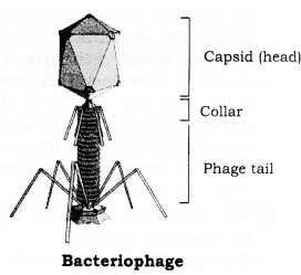
Explain the structure of Virus? Draw a labelled diagram of Bacteriophage
Answer
Viruses are non-cellular structures composed of a protein shell called capsid and a core of nucleic acids. They are so minute that they can even pass, through the microfilters and can be stored in the form of crystals. They are infectious agents, which can reproduce only in another living cell that acts as a host. They are parasitic for the purpose of the replication of their genetic material as they, themselves, do not possess the replication enzymes, etc.
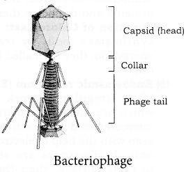
Viruses may contain DNA or RNA but, never both types of nucleic acids. Those which contain RNA as the genetic material are called retroviruses or riboviruses and there are about 400 types of RNA viruses. Those which contain DNA are called deoxy viruses. This genetic material is also called viral chromosome or viral genome which is always in the form of a small molecule. Those viruses which parasitise on bacteria are termed as bacteriophages.
Structure of Living Organisms Long Answer Type Questions
Question 31.
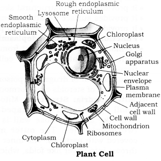
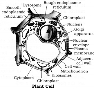
Draw a labelled diagram of a plant cell? Explain the structure and function of the following organelles? (a) Chloroplast
(b) Endoplasmic reticulum
(c) Mitochondria
(d) Nucleus
Answer
Plant cell:
Cells are comparatively larger in size. The cell wall is present. Plastids are present. Vacuoles- generally only one, large vacuole present. Dictyosomes (sub-units of Golgi body) are present. Lysosomes – either absent or very few in number. Centrosome and centrioles are absent; instead, polar caps are present.
Structure and functions:
1. Chloroplast: A chloroplast is bounded by two membranes. The inside of chloroplast is clearly marked into a colourless ground matrix, called stroma, and a membranous system called grana (singular-granum). Each granum consists of a stack of membrane-bounded, flattened sacs called thylakoids. Thylakoids contain molecules of green chlorophyll and yellow-orange carotenoid pigments, which take part in trapping solar energy. Stroma contains enzymes (for dark reactions of photosynthesis in chloroplast), DNA, RNA and ribosomes. The latter makes the chloroplasts semiautonomous. Like mitochondria, they can also synthesise some of their own proteins and duplicate themselves.
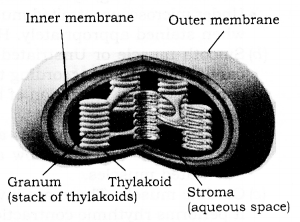
- The function of Chloroplast: Chloroplast carries out photosynthesis in plants and synthesises food, by trapping solar energy. So, they are called kitchen of the cell.
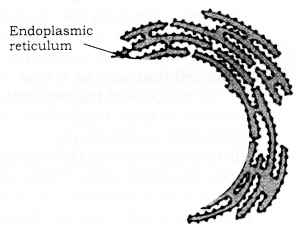
2. Endoplasmic reticulum (ER): It consists of an irregular network of channels, which are membrane-bounded and are interconnected with each other. They are seen with the help of an electron microscope only. If ribosomes are seen attached to their surface, then they are called Rough Endoplasmic Reticulum (RER). If ribosomes are not present on ER, they are called Smooth Endoplasmic Reticulum (SER).
It performs the following important functions:
- The endoplasmic reticulum acts as secretory’, storage, circulatory and nervous system of the cell.
- ER provide channels for quick transport of materials.
- It provides an ultrastructural skeletal framework of the cell.
- It provides a surface for attachment of ribosomes.
- It provides materials, lipids and proteins for biogenesis of membranes.
The function of the smooth endoplasmic reticulum is as follows:
- Formation of visual pigment from Vitamin A in retinal cells.
- Synthesis of fats inside the cells of adipose tissue.
- Detoxification of drugs and poison.
- Synthesis of steroids and hormones.
3. Mitochondria: Structure:
- Mitochondria are small spherical or rod-shaped, organelles. It is like a bag, whose wall is made up of two membranes. The outer membrane is covered with particles. The inner membrane is thrown into finger-like folds, which project into interspace called matrix. Many respiratory enzymes are present in the matrix and the inner surface of the inner membrane.
- Function: Mitochondria is the site for respiration of the cell. Enzymes control and regulate the release of energy during oxidation of food molecules in respiration. The chemical energy of food molecules is converted in the form of energy, that can be used by the cell to carry out its various activities. That is why mitochondria are called the “powerhouse” of a cell.
4. Nucleus: Robert Brown in 1831 discovered the nucleus in the cell.
Structure: The nucleus is of large size, more dense, spherical or oval and prominent organelle of the cell.
The structure of the nucleus can be divided into three parts.
- Nuclear membrane: Nucleus is enclosed by a double-layered nuclear membrane. In bacteria and blue-green algae, the nuclear membrane is not found.
- Nucleoplasm: In nucleus is present a thick jelly-like semi-fluid the nucleoplasm. In it, are observed a network of fine thread-like structures, called chromatin network. At the time of cell division, the chromatin takes the form of a thread like structures called chromosomes. The number of chromosomes is fixed for every species of animals and plants. The man has 23 pairs of chromosomes. Wheat has 21 pairs of chromosomes. The two partners of chromosomes pair are called homologous chromosomes.
- Chromosomes: Chromosomes are composed of specific proteins and a nucleic acid called Deoxy-ribose, Nucleic Acid (DNA). Genes are segments of DNA. Genes control and carry the hereditary characters.
- Nucleolus: Nucleolus contains RNA, commonly there is a single nucleolus, but two or more may be present. The nucleolus is the seat for the synthesis of cytoplasmic ribosomes. During cell division, nucleolus disappears in early stages and reappears on the same location in the daughter nuclei.
The functions of the nucleus are as follows:
- The nucleus controls the activities of the cell through its DNA molecules.
- The main function of the nuclear reticulum is participation in cell-division.
- Genes are located in a linear fashion, in chromosomes. The genes are composed of DNA. Genes control and carry the hereditary characters from one generation to another generation.
- Nucleolus assists in protein synthesis. Formation of ribosomes takes place in the nucleolus.
Question 32.
What is Mitosis? Explain the different phases of mitosis division with a labelled diagram.
Answer
Mitosis: The term Mitosis was proposed by W. Fleming-It is a type of cell division during which a cell divides into two daughter cells. Each containing the similar and same number of chromosomes as present in the parent cell.
Interphase: During this phase doubling of DNA and chromosomes takes place. Interphase is the time between the end of telophase and the beginning of the next phase.
The process of mitosis is studied in two parts:
(1) Karyokinesis
(2) Cytokinesis.
Karyokinesis: Division of the nucleus is called Karyokinesis. During this, many events take place.
- Main stages are:
(a) Prophase
(b) Metaphase
(c) Anaphase
(d) Telophase
(a) Prophase: During prophase,
- Chromosomes become very clear in the form of coiled threads.
- Chromosomes shorten and become very thick.
- These chromosomes split up longitudinally into two identical filaments called chromatids. Both the chromatids of a chromosome are attached to a centromere,
- The nuclear membrane and nucleolus dissolve away,
- In cells of flowering plants, spindle forms.
(b) Metaphase: All chromosomes arrange themselves on the equatorial plate.
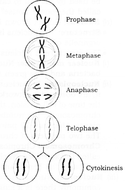
(c) Anaphase: It is the third stage of nucleus division.
- The centromeres duplicate and split. One centromere is attached to each chromatid. Separated chromatids are now called daughter chromosomes.
- Daughter chromosomes move to the opposite poles, by the contraction of the spindle fibres.
(d) Telophase:
- All the daughter chromosomes after reaching the poles uncoil and become invisible,
- The nuclear membrane is formed around the chromatin material.
- Astral rays and spindle disintegrate,
- Nucleus becomes very distinct and is known as the daughter nucleus,
- Two daughter cells are formed.
(2) Cytokinesis: Division of the cytoplasm is known as Cytokinesis. After karyokinesis, the somatic cells divide by cell plate method. In a plant cell, minute cytoplasmic vesicles filled up with substances such as calcium and magnesium in pectinate, are arranged in a line in the middle of the equatorial plane and divide the cytoplasm into two parts by progressing from middle to periphery. The cell wall is formed, due to a collection of cellulose on both sides of the plate.
A cleavage furrow appears at the equator of the animal cell and deepens progressively, until the daughter cells separate.
Question 33.
Define Tissues? Describe the simple tissues found in plants with a diagram?
Answer
Tissues: A tissue is a group or collection of similar cells, which work together to achieve a particular function and have a common origin. Blood, phloem and muscles are examples of tissues.
- Simple tissue: These are made up of only one type of cells. This type of tissue is similar in structure and function.
Simple tissues are further classified into three types:
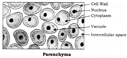
- Parenchyma
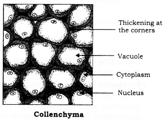
- Collenchyma
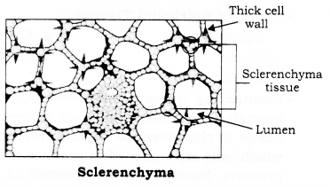
- Sclerenchyma
(1) Parenchyma: These are the simplest form of tissues with little specialisation. They have relatively unspecialised cells, with thin cell walls. They are loosely packed with inter-cellular spaces. This tissue provides support to plant, store food and allows the exchange of gases.
(2) Collenchyma: Collenchyma, being a strong and flexible tissue, has living and thick-walled cells; thickenings are present at the corners of the cells and contain cellulose and pectin; lignin is never present. Intercellular spaces are absent. Cells may be circular, oval or polygonal in shape.
A few chloroplasts may be present in the cells. Collenchyma is present in the peripheral regions of stem and leaves. Collenchyma is mainly a mechanical tissue and provides mechanical strength and elasticity to the growing stems.
(3) Sclerenchyma: Sclerenchyma is a strengthening tissue. Cells are dead and possess hard, rigid, very thick lignified walls; lignin is a waterproof material. Intercellular spaces are absent. Sclerenchyma cells are of two types-fibres which are long, narrow, pointed cells, and sclereids which are shorter, isodiametric or irregular cells; sclereids are also called stone cells or grid cells. The walls of sclerenchyma cells contain oblique thin areas, called pits. Sclerenchyma is a mechanical and protective tissue.
Question 34.
Describe the various tissues found in animals?
Answer
There are four major types of tissues in multicellular animals:
- Epithelial tissues: Protective and covering tissues.
- Connective tissue: Connecting, packing and supporting tissues.
- Muscular tissue: Contracting & relaxing tissues, carrying movements of various organs.
- Nervous tissue: Conducts impulses between brains and other organs.
Depending on the shape and function of the constituent cells.
Epithelial tissue is classified as follows:
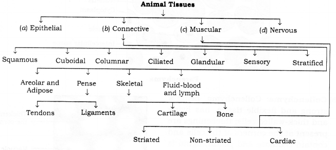
Epithelial Tissue: Epithelium forms the outer protective covering all over the body, and lies inside all the cavities such as those of the mouth, throat, stomach, intestines, windpipe and lungs. The epithelial cells lie close together with little or no intercellular substance or matrix between them. There is no blood or lymph supply. Nerve supply is present. Epithelial cells are attached to the underlying tissues by a basement membrane, which is made of a network of white, non-elostic collagen fibres. Epithelium may be one-cell thick, that is, single layered or it may be several cell thick, that is many-layered.
Types of Epithelial Tissues:
- Squamous
- Columnar
- Cuboidal
- Ciliated
- Glandular
- Sensory
- Stratified
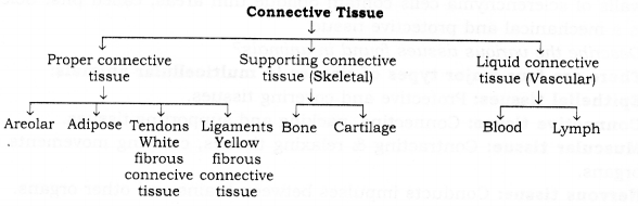
Connecting tissue: Connective tissue is the supporting and binding tissue in animals. It supports and packs different tissues and organs together. Connective tissue is characterised by the presence of a large amount of non-living matrix, in which cells are embedded. It produces the matrix, ingest bacteria and produce certain proteins. Matrix is made of semifluid, rigid or gelatinous substances. Fibres are white collagen fibres and yellow elastin fibres.
Connective tissue is of the following types:
Muscular tissue:
a. The contractile tissues are made of muscle cells, which are elongated and large sized and are so, called muscle fibres.
b. These tissues help in various types of movements of body parts and locomotion.
c. On the basis of their location, structure and function, they are of three types:
- Striated, muscles,
- Unstriated muscles,
- Cardiac muscles
(1) Striated muscles: They are attached to the bones and help in body movement and so, called skeletal or striated muscles. The striated muscle cells are long or elongated, cylindrical, unbranched and multinucleate. These muscles provide the force of locomotion and all other voluntary movements of the body.
Since the entire muscle fibres show alternate dark and light bands or stripes or striations, they are called striped muscles. These muscles work according to our will, so they are also called voluntary muscles. Their nuclei are peripheral in position. Striated muscles occur in muscles of limbs, body wall, face, neck, etc.
(2) Unstriated muscles: They occur as bundles or sheets of elongated spindle-shaped cells or fibres, with a single centrally located Eiger shaped nucleus, in the centre of cytoplasm or sarcoplasm and contractile threads called myofibrils, which run longitudinally through the cell. Since the fibrils do not bear bands or striations, hence they are smooth muscles. These muscles do not work according to our will, so, they are called involuntary muscles. Smooth muscles control the movement of food in the alimentary canal or the contraction and relaxation of blood vessels. They are also found in the iris of the eye, in the uterus, and in the bronchi of the lungs.
(3) Cardiac muscles: This type of muscle tissues are present in the muscles of the heart and are composed of branched, cylindrical and uninucleate cells. Cardiac muscles show rhythmic contraction and relaxation of the heart and help to pump and distribute blood to various parts of the body.
(d) Muscles contain special proteins called contractile proteins, which contract and relax, to cause movement.
(e) The movement of internal organs, such as heart and alimentary canal, are all caused by muscular tissues.
Nervous tissue: The cells of nervous tissue are highly specialised for being stimulated and transmitting the stimulus very rapidly from one place to another within the body. The brain, spinal cord and nerves, all are composed of the nervous tissue. The cells of this tissue are called neurons or nerve cells. Each neuron has three parts i.e. the cyton or cell body, Nucleusdendrites and the axon.
- Cyton: It is the cell body of a nerve cell that has a large central nucleus and cytoplasm, from which long, hair-like parts arise.
- Dendrite: These are the short branched fibre of neuron, which receives nerve impulses.
- Axon: It is the single long conducting fibre extending from a neuron, that transmits impulses away from the cell body,
Question 35.
Write a short note on:
(a) Vascular bundles
(b) Nervous tissue
(c) Bacteriophage
(d) Sclerenchyma
Answer
(a) Vascular bundles: They are originated from the derivatives of procambium, which itself is derived from the apical meristem. Each vascular bundle is made up of Xylem and Phloem. The cambium may be present or absent. Depending upon this, vascular bundles are of two types.
- Open Vascular bundle: In these bundles cambium is present between Xylem and phloem. For instance, dicotyledons.
- Closed vascular bundle: In these bundles cambium is absent in the vascular bundle. For instance, monocotyledons.
(b) Nervous tissue: The cells of nervous tissue are highly specialised for being stimulated and then, transmitting the stimulus very rapidly from one place to another, within the body. The brain, spinal cord and nerve, all are composed of the nervous tissue. The cells of this tissue are called nerve cells or neurons. Each neuron has three parts- the cyton or cell body, dendrites and the axon.
- Cyton: It is the cell body of a nerve cell, that has a large central nucleus and cytoplasm, from which long thin hair-like parts arise.
- Dendrite: It is the short branched fibre of neuron, which receives nerve impulses.
- Axon: It is the single long conducting fibre extending from a neuron, that transmits impulses away from the cell body.
(c) Bacteriophage: Bacteriophages can be found in all types of bacteria. Structurally, bacteriophages are almost similar to other types of viruses. They are also composed of protein and a nucleic acid but differ from other viruses in having bacteria as their host cells. It has a tadpole-like shape, with a hexagonal head and tail. The length of the head is 650 A in diameter. It has a protein coat, in which a double-stranded DNA is tightly packed. The cylindrical tail is made entirely of proteins. It has a core tube. The basal end of the tail has a hexagonal spiked end plate. At each corner of the hexagonal plate, there is a thin tail fibre. The core tube of the tail is surrounded by a contractile sheath. It is attached to the neck by a collar.
(d) Sclerenchyma:
- The cells of sclerenchymatous tissue are dead.
- They are long and narrow in appearance.
- Wals are thickened due to lignin, a chemical substance, which acts as cement and hardens them.
- Due to the presence of such strong walls, there is no internal space inside the cell.
This type of tissue is present in stems around vascular bundles in the veins of leaves and in the hard covering of seeds and nuts.
Functions:
- It is known to be the chief mechanical tissue, which makes plant hard and stiff, e.g. husk of coconut is made up of sclerenchymatous tissue.
- It provides strength and enables them to bear stress.
- It forms a protective covering around seeds and nuts. It gives rigidity, flexibility and elasticity to the plant body.
Structure of Living Organisms Additional Questions Solved
Multiple Choice Questions (MCQs)
Question 1.
The structureless fluid of cytoplasm is called as-
(A) Trophoplasm
(B) Protoplasm
(C) Hyaloplasm
(D) Metaplasm
Answer: C
Question 2.
The example of a Prokaryotic cell is –
(A) Blue-green algae.
(B) Fungi
(C) Chloroplast
(D) Animal
Answer: A
Question 3.
In which of the following DNA is found-
(A) Endoplasmic reticulum
(B) Ribosomes
(C) Chloroplast
(D) Golgi bodies
Answer: C
Question 4.
In which cell organelle respiratory enzymes are found-
(A) Cellular membrane
(B) Cell wall
(C) Mitochondria
(D) Lysosome
Answer: C
Question 5.
The organelle related to Protein synthesis is-
(A) Chloroplus
(B) Centrosome
(C) Metaplast
(D) Ribosome
Answer: D
Question 6.
The cell theory of Schleiden and Schwann stated that
(A) cells are fundamental structural units of plants and animals
(B) all cells have nuclei
(C) all cells have a nucleolus
(D) cells reproduce by mitosis and meiosis
Answer: A
Question 7.
A plant cell can be distinguished from an animal cell.
(A) By the presence of centrosome
(B) By the presence of cell wall
(C) By the presence of plasma membrane
(D) By the presence of small vacuoles
Answer: B
Question 8.
Which cell organelle is called ‘Protein Factory’ and ‘cell engine’?
(A) Golgi body
(B) Centrosome
(C) Lysosome
(D) Ribosome
Answer: D
Question 9.
In which cell organelle of the plant’s carbohydrate is synthesized, in the presence of sunlight
(A) Chloroplast
(B) Nucleus
(C) Endoplasmic reticulum
(D) Mitochondria
Answer: A
Question 10.
Which of the following is a direct division-
(A) Mitosis
(B) Meiosis
(C) Amitosis
(D) None
Answer: C
Question 11.
In which phase DNA is synthesised-
(A) Growth period II
(B) Growth period I
(C) Synthesis period
(D) Division period
Answer: C
Question 12.
A cell has 4 chromosomes. After meiotic cell divisions, the number of chromosomes in the daughter cell will be-
(A) 4
(B) 2
(C) 16
(D) 32
Answer: A
Question 13.
Synapsis occurs during-
(A) Amitosis
(B) Mitosis
(C) Meiosis
(D) None of the above
Answer: C
Question 14.
The points where two non-sister chromatids crossover are called-
(A) Chiasmata
(B) Centromere
(C) Chromomeres
(D) Chromatids
Answer:B
Question 15.
The points where two of the four chromatids cross each other are known as-
(A) Chiasmata
(B) Centromere
(C) Chromomeres
(D) Homotypic
Answer: A
Question 16.
If chromosomes are at middle part in a line, then which will be the stage of mitosis-
(A) Telophase
(B) Metaphase
(C) Prophase
(D) Anaphase
Answer: B
Question 17.
The tissue is a group of cells-
(A) Similar in origin but dissimilar in structure and function.
(B) Similar in origin, structure, and function.
(C) Similar in origin and structure but dissimilar in function.
(D) Dissimilar in structure, origin and function.
Answer: B
Question 18.
The chief function of phloem is the conduction of-
(A) food
(B) minerals
(C) water
(D) all the above
Answer: A
Question 19.
Meristematic cells are-
(A) differentiated and vacuolated
(B) mature and densely cytoplasmic
(C) mature and dead
(D) immature and living
Answer: D
Question 20.
Lateral meristem contributes-
(A) growth in thickness
(B) growth in length
(C) longitudinal growth
(D) growth in cortex
Answer: A
Structure of Living Organisms Very Short Answer Type Questions
Question 1.
Where are proteins synthesised inside the cell?
Answer
Proteins are synthesized in the ribosomes.
Question 2.
List the constituents of the plasma membrane.
Answer
The plasma membrane is made up of proteins and lipids.
Question 3.
Name two cell organelles that have their own genetic material.
Answer
Two cell organelles that have their own genetic material are mitochondria and plastids.
Question 4.
Which cell organelle is concerned with cell secretions?
Answer
Golgi bodies.
Question 5.
Why are mitochondria able to make their own proteins?
Answer
Mitochondria have their own DNA and ribosomes. Therefore, they are able to make their own proteins.
Question 6.
Which cell organelle is rich in acid, hydrolases?
Answer
Lysosome.
Question 7.
Which structure of animal cells forms the asters of the spindle?
Answer
Centrioles.
Question 8.
Write the name of different plant parts in which chloroplast, chromoplast and leucoplasts are present.
Answer
- Chloroplast-Green leaves, green stems and other green parts.
- Chromoplast-Flower (petals) and fruits.
- Leucoplast-Storage parts; such as roots, seeds, tubers etc.
Question 9.
In which stage of cell division, DNA contents get doubled?
Answer
Interphase.
Question 10.
Why Mitosis is significant?
Answer
Mitosis is significant because it produces identical cells.
Question 11.
Why does chromosomal number reduce to half in Meiosis?
Answer
This is because centromere does not divide at Metaphase-I.
Question 12.
Which is the longest stage of the first meiotic division?
Answer
The longest stage of the first meiotic division is prophase.
Question 13.
What are constituents of phloem?
Answer
Phloem is a complex tissue is made up of four types of elements:
- Sieve tube,
- Companion cells,
- Phloem fibers (bast fibers), and
- Phloem parenchyma.
Question 14.
Name the regions in which parenchyma tissue is present?
Answer
Parenchyma is a simple permanent tissue of angiospermic plants. It is present in cortex and pith of stem and roots. It is also present in the mesophyll of leaves.
Question 15.
What is the function of meristematic tissue?
Answer
Meristematic tissue functions to produce new cells, which keeps on differentiating to form specialised cells.
Question 16.
Name any two simple and two complex permanent tissues in plants.
Answer
Simple: Parenchyma and Collenchyma Complex: Xylem and Phloem.
Question 17.
Name the chief mechanical supporting tissue in plants.
Answer
Collenchyma.
Question 18.
What is tracheid?
Answer
A tracheid is a single cell, whereas trachea or vessels are tube-like bodies, formed by rows of cells.
Question 19.
Which of the following plant tissues do not possess living protoplasm at maturity?
Answer
Sclerenchyma.
Question 20.
Name the following:
- Multinucleate muscle fibre
- Spindle-shaped muscle fibre
Answer
- Skeletal muscle fibre
- Smooth muscle fibre.
Structure of Living Organisms Short Answer Type Questions
Question 1.
Why is a cell called the structural and functional unit of life?
Answer
All living organisms are made up of cells. Thus, the cell is the structural unit of life. Each living cell has the capacity to perform certain basic functions, that are characteristics of all living forms. Each cell acquired distinct structure and function, due to the organisation of its membrane and cytoplasmic organelles in a specific way. Each kind of cell organelle performs a special function, such as making new materials in the cell (e.g. chloroplast, ribosomes), clearing up the waste materials from the cells (e.g. lysosomes), utilisation of oxygen, oxidation of food and energy production (e.g. mitochondria), movement (microtubules containing spindle, cilia flagella), etc. A cell is able to live and perform all its functions because of these organelles. These organelles together constitute the basic unit called ‘cell’.
Question 2.
Why is plasma membrane, called a selectively permeable membrane?
Answer
Plasma membrane allows movement of only selected molecules across it, so it is called selectively permeable membrane. For instance, it permits the entry of gases through diffusion and water through osmosis. Larger molecules may pass through the plasma membrane by an active process. The plasma membrane is impermeable to certain other materials. Therefore, it is selectively permeable.
Question 3.
Which organelle is known as the powerhouse of the cell? Why.
Answer
Mitochondria are known as the powerhouse of the cell because the energy required for various life activities is released by mitochondria in the form of ATP molecules. ATP is known as the energy currency of the cell and is used as cellular fuel. Energy stored in ATP is used to carry out energy-requiring activities of the cell, such as photosynthesis, protein synthesis and muscle contraction.
Question 4.
Why do plant cells possess largely sized vacuole?
Answer
Vacuole of a plant cell has to be large because it takes part in maintaining the water level of the cell, i.e. entry and exit of water. In addition, vacuole helps in-
(a) Storage- It store sugars, amino acids, organic acids, salts and some proteins.
(b) Cellular wastes- They are dumped in the vacuole.
(c) Turgidity-The vacuole contains cell sap, which provides turgidity to the cells.
(d) Absorption of water- Vacuole contains an osmotic concentration, required for absorption of water.
Question 5.
What are viruses? Why do they not show any characteristics of life?
Answer
Cell organelles which perform specific functions for the cell are enclosed by membranes. But, viruses do not have any membrane. So viruses do not show any characteristics of life on its own. Outside its host cell, a virus is completely inert. They become active only when they enter in some living body and uses its cell machinery.
Question 6.
Where are chromosomes located? What are they composed of? What is chromatin material and how does it change, just before the cell divides?
Answer
Chromosomes are located in the nucleus of plant and animal cells. They are composed of DNA and protein. Chromatin material is entangled mass of thread-like structures. Chromatin material gets organised into chromosomes, just before the cell divides.
Question 7.
How is a prokaryotic cell different from a eukaryotic cell?
Answer
Prokaryotic Cell:
- Size of the cell is generally small (1-10 m).
- The nuclear region is poorly defined, due to the absence of nuclear membrane, known as a nucleoid.
- It contains a single chromosome.
- The nucleolus is absent.
- Membrane-bound cell organelles are absent.
Eukaryotic Cell:
- Size of the cell is generally large (50 – 100 m)
- The nuclear region is well-defined and surrounded by a nuclear membrane.
- It contains more than one chromosome.
- The nucleolus is present.
- Cell organelles such as mitochondria, plastids, endoplasmic reticulum, golgi apparatus, lysosomes, peroxisomes, etc. are present.
Question 8.
What are lysosomes, peroxisomes and centrosomes? Write their functions.
Answer
Lysosomes: They are single- membrane small vesicular structures, found in the cytoplasm of all eukaryotic cells, except mammalian RBC’s. They contain enzymes and are formed by Golgi apparatus.
Functions: They are involved in intracellular digestion of foreign food or microbes and are also involved in autolysis or self-digestion of cells, after their death.
Peroxisomes: They are found in photosynthetic cells of plants, liver and kidney cells of the vertebrates and contain two types of oxidative enzymes: oxidase and catalase, bounded by a unit membrane.
Functions: These are involved in the removal of toxic substances, by oxidative reactions. In plant cells, these also help in photorespiration.
Centrosome: A centrosome is a light microscopic organelle, formed of two dark, coloured granules called centrioles, surrounded by a transparent cytoplasmic area called centrosphere. It lies near the nucleus and is commonly called the call centre.
Functions: Centrosome helps in cell division, in animal cells. They also help in the formation of cilia and flagella of the cells.
Question 9.
Describe the structure and function of the Golgi apparatus?
Answer
Following are the structure and functions of Golgi apparatus:
First described by Camillo Golgi, and also known as Golgi body or Golgi complex, it consists of smooth, flattened, membrane-bound (double- walled), sac-like structures called cisternae.
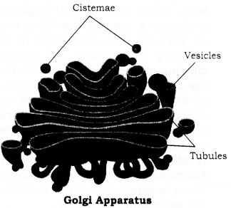
Golgi Apparatus
- The cisternae are usually placed one above the other (stacked together in parallel rows.
- The Golgi apparatus is frequently surrounded by vesicles, which are discharged from the cisternae.
- When present in plants in the form of subunits, golgi apparatus is called dictyosome.
Functions:
- It is secretory organelle of the cell.
- It packages materials synthesised in the cell and transports it.
- It is involved in the formation of lysosomes.
- In some cases, complex sugars are made from simple sugars in this organelle.
Question 10.
Explain cell division?
Answer
Cell Division: The German biologist Walter Flemming was the first, who observe the process of cell division, and gave it the term mitosis. A cell division is a process by which a single cell multiplies in number. A growing cell undergoes interphase (a period of non-apparent division) and the period of division. In interphase, cell doubles in size and chromosomes duplicate. Therefore, cell division can be considered a process of ensuring exact duplication of cellular information and its equal distribution to the two daughter cells. Thus, the two type of cell divisions observed, are mitosis and meiosis.
Question 11.
In which phase, gene exchange between chromatids take place?
Answer
The process of gene exchange takes place in the prophase I of meiosis. In the beginning of prophase I, chromosomes appear as long thread like structure and show bead-like thickenings at regular intervals. The homologous chromosomes form pairs. This phenomenon of chromosomal pairing is called synapsis of the chromosomes. The paired chromosomes are identical, one coming from the father, through his sperm and the other from mother, through her ovum. Pairing is remarkably exact and specific. It takes place to point to point. The two homologues do not fuse during pairing but remain separated. The paired chromosomes become short and thick and contain four chromatids.
Each chromatid at this stage has its own centromere, resulting in four centromeres. These chromatids are called sister chromatids. Now, two of the chromatids (one paternal and one maternal) of the homologous, exchange segments. The process is called crossing over.
Question 12.
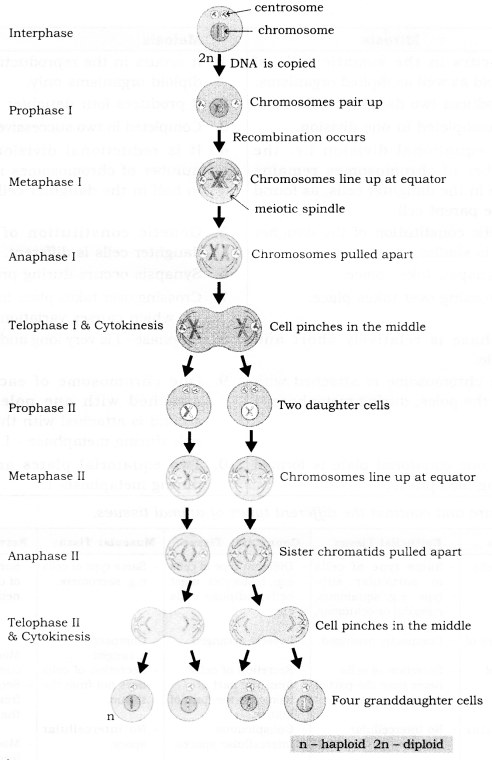
Describe Meiosis?
Answer
Meiosis is an indirect type of cell division, which occurs at the time of gamete formation. It involves two successive divisions, rapidly occurring one after the other and results in the formation of four daughter cells, with half the number of chromosomes as compared to the parent cell. Such cells are called haploid cells (n), whereas parent cells are diploid (2 n). The 1st division of meiosis reduce the chromosomal number to half and is, therefore, called the reductional division. It produces two haploid cells from the parent diploid cell. The Ilnd division of meiosis is similar to mitosis, as the chromosomal number of the haploid cells produced in the reductional division above, remains unchanged. Four haploid cells result at the end of meiosis.
Question 13.
Explain with a diagram, the objective of amitosis in bacteria?
Answer
The objective of amitosis in bacteria is to increase progeny and propagation of generation.
Amitosis: The nuclear material containing a circular chromosome, elongates. A constriction develops in the middle portion of a chromosome. When constriction increases the circular chromosome, it divides into two halves. In the beginning, the chromosome is attached to the cell wall. But as the cell increases in size, the two halves of chromosome separate. After that, the constriction develops into the cell wall. When constriction increases, the bacterial cell divides into two daughter cells.
Question 14.
Explain centromere, centriole and chaismata.
Answer
Centromere: Chromosomes are mostly of the same length and thickness, but at one or more places they are constructed. These places are known as primary constrictions or centromere. Usually, one chromosome possesses only one centromere, but some of them may have more than one centromere.
Centriole: It is a cell organelle of animal cells. It takes part in the spindle formation, during nuclear division. Each animal cell normally possesses a pair of centrioles, lying with their axes at right angles to each other.
Charismata: Charismata is the region on homologous chromatids where the exchange of genetic material (crossing over) takes place.
Question 15.
What are the functions of areolar tissue?
Answer
Main functions of areolar tissue are as follows:
- These tissues fill the space inside the organs.
- It provides support, elasticity and strength to the body parts.
- Aerolar tissue binds different tissues together, e.g., the skin with underlying parts, blood vessels and neurons to other body parts.
- It helps in repair of tissues and healing of wounds.
- It helps in fighting foreign antigens and toxins.
Question 16.
How are simple tissues different from complex tissues, in plants?
Answer
Simple tissues are made up of only one type of cells, which look like each other. On the other hand, complex tissues are made up of more than one type of cells. Parenchyma, collenchyma and sclerenchyma are examples of simple plant tissue, whereas xylem and phloem are examples of a complex tissue. Simple tissues are protective and supportive in function. Complex tissues conduct water, minerals, and nutrients from roots and leaves to different parts of the plant.
Question 17.
What is the role of the epidermis in plants?
Answer
Epidermis is a protective layer of tissue in angiospermic plants. It provides protection to underlying tissues. Epidermis forms the outer covering of various plant organs, such as roots, stem, leaves and flowers and remains in direct contact with the environment. Any substance, whether solid, liquid or gas can enter into the plant or move outside, only after passing through this layer. Epidermis helps in absorption, secretion, excretion, gaseous exchange and transpiration. It helps in preventing the entry of pathogens.
Question 18.
How do cardiac muscles resemble both striated and smooth muscle fibres?
Answer
Similarities with striated muscles:
Both are cylindrical in shape, highly vascular and have alternate dark and light bands. Similarities with smooth muscles: Both are smallsized, uni-nucleated and involuntary.
Question 19.
What are mast cells? What is their function?
Answer
Mast cells are oval-shaped cells of areolar connective tissue, having dense granules in their cytoplasm. These secrete matrix of connective tissue, heparin and histamine.
Question 20.
Describe blood as connective tissue.
Answer
Blood is a fluid connective tissue. In blood, the cells move in a fluid matrix called blood plasma. The cells in blood are different from the other connective tissue cells, both in structure and functions. Blood plasma is a straw coloured, slightly alkaline fluid. Blood plasma contains cells called blood corpuscles and blood platelets. Blood cells are of two types-Erythrocytes and Leucocytes. Blood circulates within the blood vessels throughout the body and thus, connect every part of the body.
Structure of Living Organisms Long Answer Type Questions
Question 1.
Name the three major functional regions of cells. Briefly mention the component of each and explain the function of each. Draw a labelled diagram of a plant cell.
Answer
All cells vary in their shape, size and activities and have three major functional regions, viz, plasma membrane, nucleus and cytoplasm.
Plasma Membrane or Cell Membrane: This is the outermost covering of the cell, that separates the contents of the cell from its external environment. The plasma membrane is a living, thin, delicate, elastic, a selectively permeable membrane made up of proteins and lipids and is present in both plant and animal cells.
Functions of Plasma Membrane:
- It gives definite shape to the cell.
- It separates the contents of a cell from its surrounding medium.
- It provides a mechanical barrier, for the protection of the internal contents of the cell.
- It is a selectively permeable membrane.
- It regulates the movement of ions, in and out of the cell.
Nucleus: Robert Brown in 1831 discovered the nucleus in the cell. The nucleus is the largest cell structure. It is a spherical or oval, prominent structure, usually located in the centre of the cell. Nucleus has the following important parts:
Nuclear membrane: It is a double-layered membrane, which separates the nucleus from the cytoplasm. It has pores called nuclear pores, which allow the transfer of material from the nucleus to the cytoplasm. Nucleoplasm: It is a homogeneous and granular dense fluid, present inside the nucleus, in which chromatin and nucleolus are suspended.
Chromatin material: It consists of a long, coiled network of thread-like structures. The chromatin material is made up of deoxyribonucleic acid (DNA), which is responsible for storing and transmitting the hereditary information, from one generation to the other. It condenses into compact rod-like bodies, called chromosomes, at the time of cell division.
Nucleolus: It is more or less round in structure, found inside the nucleus. The nucleolus contains RNA (ribonucleic acid) and proteins. RNA is helpful in protein synthesis in the cytoplasm.
Functions of Nucleus:
The nucleus controls all the metabolic activities of the cell.
- It regulates the cell cycle.
- It is concerned with the transmission of hereditary traits from the parent to offsprings.
- Cytoplasm: It is the fluid content of the cell, which occurs between the plasma membrane and the nuclear envelope. It contains various cell organelles, which perform different functions of the cell.
Functions of Cytoplasm:
- Cytoplasm helps in exchange of materials between cell organelles.
- It acts as a store of vital chemicals such as amino acids, glucose, vitamins, ions, etc.
- It is the site of certain metabolic pathways, such as glycolysis, synthesis of fatty acids, nucleotides. Some amino acids are also synthesised in the cytoplasm.
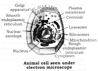
Question 2.
Draw a labelled diagram of an animal cell. Describe the structure and give the name of four cell organelles.
(1) Mitochondria
(2) Ribosomes
(3) Lysosomes
(4) Endoplasmic reticulum
Answer
(1) Mitochondria: These are small rod-shaped organelles. Each mitochondrion is bounded by a double membrane. The outer membrane provides a smooth, uninterrupted boundary to the mitochondrion.
The inner membrane is raised up into a large number of finger-like projections, called cristae, towards the interior of the mitochondrion. The interior is known as matrix. Matrix is filled with semi-liquid fluid. In a matrix and on the inner side of it, many enzymes are present, which control and regulate the release of energy during oxidation of food molecules, in respiration.
Function: Mitochondria is the site of respiration. Energy is released during oxidation of food, in the form of chemical energy, stored as ATP molecule(s). This energy can be used by the cell to carry out its various activities. That is why mitochondria are the powerhouse of a cell.
(2) Ribosomes: Ribosomes are small, dense, rounded and granular particles, made up of RNA, proteins, and remain with the membrane of the endoplasmic reticulum. They may occur free, in cytoplasm also.
Function: The main function of ribosomes is to synthesise protein and it regulates the protein synthesis.
(3) Lysosomes: Lysosomes are rough, spherical sac-like structures, bounded by a single membrane. They are filled with dense liquid. The liquid mainly consists of enzymes, which help in the digestion of food molecules.
Functions:
- They control the digestion of cells.
- They help in the digestion of dead cells and disintegration cells.
- They are more in number, in white blood corpuscles. That is why they are capable of digesting the microbes or bacteria.
(4) Endoplasmic reticulum: They were discovered by Keith Porter. These organelles are found in eukaryotic cells. These organelles are not found in prokaryotic cells and in red blood corpuscles of mammals.
These are of two types:
- Smooth endoplasmic reticulum: Ribosomes are not found on their outer surface. These are found mainly in those cells which synthesise and store carbohydrates, fats, steroid hormones and other products, devoid of proteins.
- Rough endoplasmic reticulum: Ribosomes are stuck firmly on their outer surface.
Functions of endoplasmic reticulum:
- They transport different soluble materials inside the cell.
- They are helpful in the synthesis of protein and steroid hormones.
- They manufacture the nuclear membrane and golgi bodies.
Question 3.
Give briefly the difference between plant and animal cell
Answer
| Plant Cell | Animal Cell | |
| 1. | A plant cell is comparatively large in size. | The animal cell is usually smaller in size. |
| 2. | The plasma membrane is surrounded by a rigid, thick and non-living cell wall made up of cellulose. | The animal cell is only enclosed by living plasma membrane, and lack cell wall. |
| 3. | There are one or two large vacuoles, in which cell sap is enclosed by tonoplast. | Cytoplasm contains many smaller vacuoles, which are distributed in the whole cytoplasm. |
| 4. | Plastids (chloroplast and chromoplast) are very common in a plant cell. | Plastids are absent in an animal cell, except in Euglena. |
| 5. | Plant cell lack centrioles. Instead, two small clear areas called polar caps are present. | Animal cell posses centrosome with one or two centrioles. |
| 6. | Contact between plant cells is through plasma desmatic. | Contact between animal cells is through desmosome. |
| 7. | A plant cell has several subunits of less developed golgi bodies called dictyosomes. | The animal cell has permanent and highly complex golgi bodies, near the nucleus. |
| 8. | Microvilli are absent on the surface of plant cells. | Microvilli are present on the surface of animal cells. |
| 9. | Stored food is starch, in them. | Stored food is glycogen. |
| 10. | A plant cell is rigid. | The animal cell is flexible. |
Question 4.
Describe meiosis. Draw labelled diagram of any two phases.
Answer
The term ‘Meiosis’ was proposed by Farmer and Moore. Meiosis is important for organisms which reproduce sexually. It is a special type of cell division, in which cell undergoes two successive divisions, producing four daughter cells, each containing half the number of chromosomes as compared to the parent cell.
The reduction takes place because the gametes are diploid (2n). They will produce a zygote after fertilisation, with double the number of diploid chromosomes (4n). To avoid this, reduction takes place and cell become haploid from diploid, before fertilisation. So, after the fusion of gametes, the zygote has 2n-diploid chromosome number. Meiosis involves two separate cell divisions, called Meiosis I and Meiosis II.
(1) Meiosis I:
Reductional division: Meiosis I results in a reduction of chromosomes to half with the formation of two haploid cells. It takes place in four phases, i.e., prophase, metaphase anaphase and telophase.
- Prophase-I. In this phase, the nucleus increases. Centrosome separates and moves toward the poles. Chromatin network converts into chromosomes. Chromosomes are present in the form of fine threads. Attraction occurs in homologous chromosomes which arrange in pairs. This is known as synapsis or pairing. The two nonsister homologous chromatids break apart (separate) at corresponding points, due to repulsion. They remain in contact with each other at many points, which are termed as chiasmata. By the formation of chiasmata a piece of a chromatid, bearing genes get transferred from one to another chromatid and vice versa. It is known as a crossover.
- Metaphase I- Centrioles form a spindle. The pairs of homologous chromosomes arrange on the equatorial plate in such a way that centromeres are towards the poles, but their arms are towards mid-line.
- Anaphase I- The two partners of homologous chromosomes completely separate from each other and start migrating towards the opposite poles of the spindle. In this way, two sets are formed. Each set is on a pole. Now, the number of chromosomes becomes haploid.
- Telophase I– Nuclear membrane is reappears. Thus, a nucleus is reformed at each pole. Nucleolus also again appears. The division of cytoplasm (cytokinesis) by cleavage, results in the two haploid daughter cells.
(2) Meiosis II:
Equational Division: The two daughter cells then undergo Meiosis II which is exactly like mitosis. Meiosis II, is an equational division, resulting in four daughter cells with half the number of chromosomes.
- This is also completed in two stages.
(A)Kaiyokinesis.
(B) Cytokinesis.
(A) Karyokinesis-
In this, four stages take place:
(1) Prophase II
(2) Metaphase II
(3) Anaphase II
(4) Telophase II
(1) Prophase II- Two asters are formed, moving towards the opposite poles. Spindle fibres develop chromosomes, which get attached to the spindle fibre.
(2) Metaphase II- Chromosomes get arranged anged on the equatorial plane and the sister centromeres separate. Each chromosome produces two daughter chromosomes.
(3) Anaphase II- Centromere of each chromosome splits into two daughter centromeres and one chromatid of chromosome attached with one daughter centromere and other with the second centromere, now known as daughter chromosomes. Two daughter chromotids move towards, the opposite poles.
(4) Telophase II- A11 of the four nuclei has one chromatid and each nucleus has a haploid number of chromosomes, as well as DNA, as compared to the parent cell.
(B) Cytokinesis-
The karyokinesis of the second meiotic division is followed by cytokinesis. It starts at anaphase II and completed by the end of Telophase II. This process is similar to mitosis.
Question 5.
Write the difference between Mitosis and Meiosis in tabular form?
Answer
| Mitosis | Meiosis |
| 1. It occurs in the somatic cells of haploid as well as diploid organisms. | 1. It occurs in the reproductive cells of diploid organisms only. |
| 2. It produces two daughter cells. | 2. It produces four daughter cells. |
| 3. It is completed in one division. | 3. Completed in two successive divisions. |
| 4. It is equational division i.e. the number of chromosomes remains the same in the daughter cells, as found in the parent cell. | 4. It is reductional division i.e. the number of chromosomes is reduced to half in the daughter cells. |
| 5. The genetic constitution of the daughter cells is similar. | 5. Genetic constitution of the four daughter cells is different. |
| 6. No synapsis takes place. | 6. Synapsis occurs during prophase-I. |
| 7. No crossing over takes place. | 7. Crossing over takes place in prophase – I, which causes variations. |
| 8. Prophase is relatively short and simple. | 8. Prophase -1 is very long and elaborate. |
| 9. Each chromosome is attached with both the poles, during metaphase. | 9. One chromosome of each pair is attached with one pole and the second is attached to the opposite pole during metaphase – I. |
| 10. Only one equatorial plate is formed during metaphase. | 10. Two equatorial plates are formed during metaphase- I. |
Question 6.
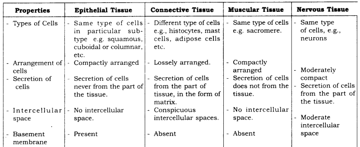
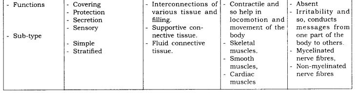
Compare and contrast the different types of animal tissues
Answer
Question 7.
In brief, state what happens when:
(a) Dry apricots are left for some time in pure water and later transferred to the sugar solution.
(b) A red blood cell is kept in concentrated saline solution?
(c) Rheo leaves are boiled in water first and then a drop of sugar syrup is put on it?
(d) The plasma -membrane of a cell breaks down?
(e) Golgi apparatus is removed from the cell?
Answer
(a) Dry apricots when left in pure water, will gain water due to endosmosis, and swell. When placed in a concentrated sugar solution, they lose water due to exosmosis and will shrink.
(b) If an RBC is kept in concentrated saline solution, it will lose water and shrink.
(c) Boiling of Rheo leaves in water kills the cells in the plasma membrane. When a drop of sugar syrup is put on it, the process of plasmolysis will not occur, because only the living cells can absorb water by osmosis, dead cells are not able to do so.
(d) If plasma membrane of a cell breaks down:
- Shapes of the cell will be affected.
- Cell components of that cell may get damaged.
- Harmful substances may enter the cell.
- Useful substances may flow out of the cell.
(e) If the Golgi apparatus is removed from the cell:
- Secretion process of the cell will be stopped.
- Packaging and transportation of useful material will be stopped.
- There will be no formation and repair of broken membranes, lysosomes, plasma membrane etc. No removal of dead cell components will take place.
- Acrosome formation in sperms will not take place, so it will not be able to enter the egg.
We hope the given RBSE Solutions for Class 9 Science Chapter 6 Structure of Living Organisms will help you. If you have any query regarding Rajasthan Board RBSE Class 9 Science Chapter 6 Structure of Living Organisms, drop a comment below and we will get back to you at the earliest.